
Page 28 - AN-3-3
P. 28
Advanced Neurology Tau pathology in murine TBI
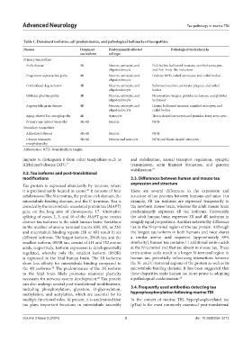
Table 1. Dominant isoforms, cell predominance, and pathological hallmarks of tauopathies
Disease Dominant Predominantly affected Pathological tau hallmarks
tau isoform cell type
Primary tauopathies
Pick’s disease 3R Neuron, astrocyte, and Pick bodies, ballooned neurons, ramified astrocytes,
oligodendrocyte and Pick-body-like inclusions
Progressive supranuclear palsy 4R Neuron, astrocyte, and Globose NFTs, tufted astrocytes, and coiled bodies
oligodendrocyte
Corticobasal degeneration 4R Neuron, astrocyte, and Ballooned neurons, astrocytic plaques, and coiled
oligodendrocyte bodies
Globular glial tauopathy 4R Neuron, astrocyte, and Neuronal pre-tangles, globular inclusions, and globular
oligodendrocyte inclusions
Argyrophilic grain disease 4R Neuron, astrocyte, and Grains, ballooned neurons, ramified astrocytes, and
oligodendrocyte coiled bodies
Aging-related Tau astrogliopathy 4R Astrocyte Thorn-shaped astrocytes and granular-fuzzy astrocytes
Primary age-related tauopathy 3R+4R Neuron NFTs
Secondary tauopathies
Alzheimer’s disease 3R+4R Neuron NFTs
Chronic traumatic 3R+4R Neuron and astrocyte NFTs and thorn-shaped astrocytes
encephalopathy
Abbreviation: NFTs: Neurofibrillary tangles.
impacts to distinguish it from other tauopathies such as and stabilization, axonal transport regulation, synaptic
Alzheimer’s disease (AD). 27 transmission, actin filament formation, and genome
stabilization. 24
3.2. Tau isoforms and post-translational
modifications 3.3. Differences between human and mouse tau
Tau protein is expressed abundantly by neurons, where expression and structure
it is predominantly located in axons. It consists of four There are several differences in the expression and
22
subdivisions: The N terminus, the proline-rich domain, the structure of tau proteins between humans and mice. For
microtubule-binding domain, and the C terminus. Tau is example, 3R tau isoforms are expressed temporarily in
encoded by the microtubule-associated protein tau (MAPT) the newborn mouse brain, whereas the adult mouse brain
gene on the long arm of chromosome 17. Alternative predominantly expresses 4R tau isoforms. Conversely,
splicing of exons 2, 3, and 10 of the MAPT gene creates the adult human brain expresses 3R and 4R isoforms in
distinct tau isoforms in the adult human brain. Variations roughly equal proportions. Another noteworthy difference
in the number of amino terminal inserts (0N, 1N, or 2N) lies in the N-terminal region of the tau protein. Although
and microtubule binding repeats (3R or 4R) result in six the longest tau isoform in both humans and mice shares
different isoforms. The largest isoform, 2N4R tau, and the a similar amino acid sequence (approximately 89%
smallest isoform, 0N3R tau, consist of 411 and 352 amino similarity), human tau contains 11 additional amino acids
acids, respectively. Isoform expression is developmentally at the N-terminal end that are absent in mouse tau. These
regulated, whereby only the smallest isoform (0N3R) extra amino acids result in a longer N-terminal region in
is expressed in the fetal human brain. The 3R isoforms human tau, potentially influencing interactions between
show less affinity for microtubule binding compared to the N- and C-terminal regions of the protein as well as its
the 4R isoforms. The predominance of the 3R isoform microtubule-binding domain. It has been suggested that
28
in the fetal brain likely promotes neuronal plasticity these disparities make human tau more prone to adopting
necessary for nervous system development. Tau protein a pathological conformation. 29
24
can also undergo several post-translational modifications,
including phosphorylation, glycation, O-glycosylation, 3.4. Frequently used antibodies detecting tau
methylation, and acetylation, which are essential for its hyperphosphorylation following murine TBI
multiple functional roles. At present, it is understood that In the context of murine TBI, hyperphosphorylated tau
tau plays important functions in microtubule assembly (pTau) is the most commonly examined post-translational
Volume 3 Issue 3 (2024) 3 doi: 10.36922/an.3213