
Page 31 - AN-3-3
P. 31
Advanced Neurology Tau pathology in murine TBI
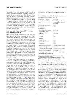
contributes to a less stable and more flexible cytoskeleton, Table 3. Murine TBI tau pathology compared to human TBI/
thereby promoting neuronal plasticity during these CTE
stages. In addition, increased Tau phosphorylation Characteristic tau lesions in CTE Murine TBI models
24
has been observed in the brains of sleeping adults and
hibernating mammals. Furthermore, widespread and pTau immunoreactivity in neurons + (33, 36)
75
reversible hyperphosphorylation of Tau has been shown Astrocytic pTau deposition + (33, 81, 85)
to occur in response to multiple metabolic stresses such pTau lesions at the depth of a cortical Cannot be replicated due to
as glucose deprivation, starvation, exposure to cold, sulcus the lisencephalic nature of the
acute immobilization, and seizures. 76-79 Further study is murine brain
required to delineate whether and to what extent pTau pTau lesions around capillaries + (33)
may promote neuroprotective and neurorestorative Subpial pTau lesions + (33)
processes following TBI. pTau pathology in
Cerebral cortex (preferentially + (33, 81, 85)
3.6. Temporal evolution of pTau differs between in the superficial layers)
brain regions following TBI
Hippocampus + (36, 81, 85)
Using enzyme-linked immunoassay, pTau has been Entorhinal cortex _
detected within the ipsilateral cortex as early as 3 h after Amygdala + (81)
murine blast TBI. However, relatively little is known about Thalamus -
80
the temporal evolution of pTau after closed-head TBI,
as most studies assessed pTau at only a single timepoint Mamillary body + (33)
(typically 1 month) after TBI, and data from the few time- Cerebellar dentate nucleus + (81)
course studies have been conflicting. 33,36,49,80-82 For instance, Tau oligomers + (94)
two studies indicated a continued rise in pTau from 3 h 3R Tau isoforms in tau oligomers - (In non-transgenic models)
to 6 months after rTBI. 36,81 In contrast, another study Insoluble tau aggregates + (36, 94, 95)
reported no change in pTau levels between 1 and 6 months Paired helical filaments and NFTs - (In non-transgenic models)
post-rTBI. In addition, three studies observed transient Trans/cis pTau transformation + (36, 49, 50) +
33
pTau with a peak at 1 month but found no significant
expression by 4 – 6 months after TBI. 49,60,73 Notably, two of Secondary spreading of Tau Not conclusive (36, 93-95)
aggregates from the initial injury site
these studies employed a single TBI paradigm, suggesting
that persistent pTau accumulation is more likely to occur Notes: “+” denotes histopathological features present in murine
traumatic brain injury (TBI) models.
after rTBI in mice. “-” denotes histopathological features absent in murine TBI models.
Cellular and spatial distribution of tau pathology Abbreviations: CTE: Chronic traumatic encephalopathy;
following human TBI has been well characterized and used NFTs: Neurofibrillary tangles: pTau: Phosphorylated Tau.
as diagnostic criteria for CTE, whereby pTau aggregates to axonal injury. 86,87 Indeed, it has been shown that the
in neurons, perivascular lesions at the depth of a cortical subpial cerebral cortex lining the superficial longitudinal
sulcus, deep in the parenchyma, and not restricted fissure represents an area of frequent pTau accumulation. 33
to the subpial and superficial sulcus are considered
characteristic. 27,83 Investigators studying the impact of In addition to the presence of pTau within neurons,
TBI on pTau formation should consider that the temporal multiple studies also found pTau within astrocytes after
evolution of pTau differs between brain regions post-injury. murine closed-skull TBI. 33,81,85 Interestingly, pTau-positive
For example, the earliest occurrence of pTau-positive cells astrocytes were predominantly localized in the superficial
33
has been reported in the corpus callosum, 33,84 with later cortex and around capillaries, raising the possibility that
involvement of the cerebral cortex, subcortical nuclei, 33,81,85 astrocytic pTau expression is a result of stretch injury to
and finally, the hippocampus. 36,81,82 (Table 3). The reasons the glia limitans, the thin line of astrocytic foot processes
for the difference in the spatial distribution of pTau may associated with the parenchymal basal lamina at the
be reflective of trauma biomechanics, tissue-specific cortical surface and the pericapillary space. As noted
88
vulnerability to injury, as well as the cellular response to above, dysregulation of aquaporin-4, which is particularly
injury. Computational models of biomechanical forces on prevalent in astrocytic membranes at the blood–brain
the brain from TBI indicated that cerebral areas with a interfaces, may lead to decreased pTau clearance through
69
change in morphology (such as at the site of sulci and the the glymphatic system. Conversely, astrocytes have
superior longitudinal fissure) represent locations of high been shown to take up pTau from the extracellular space
stress and strain and thus bear the greatest susceptibility through receptor-mediated endocytosis. 89-91
Volume 3 Issue 3 (2024) 6 doi: 10.36922/an.3213