
Page 30 - AN-3-3
P. 30
Advanced Neurology Tau pathology in murine TBI
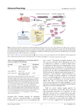
Figure 2. Potential mechanisms implicated in tau pathology following murine traumatic brain injury (TBI). Traumatic brain injury causes a cascade of
events that ultimately lead to axonal injury. This process, along with the dysregulation of tau kinases and phosphatases, promotes the hyperphosphorylation
of tau protein. Hyperphosphorylated Tau (pTau) loses its ability to bind and stabilize microtubules. Disruption of astrocyte aquaporin-4 polarity impairs
pTau clearance through the glymphatic system. Pro-inflammatory cytokines and inflammatory signals produced by activated microglia and impaired
microglial phagocytosis of pTau accentuate pTau accumulation.
Notes: * Refer to Table 3 for a list of kinases/phosphatases, ► Sarm1 activation causes axonal degeneration after TBI. Inhibition of the prodegenerative
Sarm1 pathway attenuates axonal injury and attenuates pTau accumulation, ┴ :Inhibition, **: Not shown in the mouse TBI models
70
Table 2. Tau kinases/phosphatases in AD, human TBI/CTE, brain injury. TBI-induced microglial activation may
and mouse models of TBI pathology either promote deleterious effects through the release of
pro-inflammatory cytokines or exhibit beneficial effects
Pathology Kinases Phosphatases by suppressing inflammation and enhancing tissue repair.
AD GSK3β , JNK p38 , ERK , PP1, PP2A, In particular, the pro-inflammatory M1 phenotype has
‡
‡
†
†
†
†
†
†
PKA , CamKII , c-Abl , PP2B , PP5,
CK1, CDK5, PKN, Fyn PTEN been associated with post-traumatic accumulation of pTau
‡
Human TBI/CTE GSK3β , JNK ‡ - in addition to the release of neuroinflammatory factors,
inhibition of neurite outgrowth, and neuronal apoptosis.
60,71
Murine TBI models GSK3β , JNK , p38 , ERK , PP2B † Attenuation of post-traumatic neuroinflammation, for
†
‡
‡
†
PKA , CamKII , c-Abl †
†
†
example, through enhancing endogenous cannabinoids
Notes: Indicates kinases/phosphatases that have been shown such as 2-arachidonoylglycerol (2-AG), has been shown
†
deregulated in human Alzheimer’s disease (AD) and murine traumatic
brain injury (TBI) models, Indicates kinases/phosphatases that have to decrease pTau accumulation and attenuate behavioral
‡
been shown deregulated in human AD, human TBI, and murine TBI. deficits after TBI. 72,73 Furthermore, genetic ablation of
Abbreviations: CaMKII: Ca2+/CalModulin-dependent protein kinase Sarm1 (sterile alpha and TIR motif containing 1), a key
II; c-Ab: c Abelson tyrosine kinase; CDK5: Cyclin-dependent kinase-5; regulator of axon degeneration, mitigated both microgliosis
CK1: Casein kinase 1; CTE: Chronic traumatic encephalopathy; and pTau accumulation following rTBI. 74
ERK: Extracellular receptor kinase; GSK3: Glycogen synthase kinase-3;
JNK: c-Jun N-terminal kinase; PKA: cAMP-dependent protein kinase; While the current evidence largely supports the
P38: p38 mitogen-activated protein kinase; PP1: Protein phosphatase-1; idea that the formation of pTau after TBI is associated
PP2A: Protein phosphatase-2A; PP2B: Protein phosphatase-2B;
PP5: Protein phosphatase-5; PTEN: Phosphatase and tensin homolog with pathological processes, it is important to note
deleted on chromosome 1. that pTau may also serve a neuroprotective role under
different circumstances. For instance, during embryonic
decreased pTau clearance through the glymphatic development and in the developing human brain, pTau
system. Moreover, neuroinflammation is increasingly levels are comparable to those observed in adults with
69
recognized for its important role in pTau pathology after AD. It has been suggested that tau phosphorylation
24
Volume 3 Issue 3 (2024) 5 doi: 10.36922/an.3213