
Page 29 - AN-3-3
P. 29
Advanced Neurology Tau pathology in murine TBI
modified form of tau protein. There is significant overlap in 3.5. Mechanisms implicated in tau phosphorylation
the phosphorylation sites of human and mouse tau, and there following mouse TBI
are at least 85 distinct identified phosphorylation sites. 30-32 Several lines of data indicate that TBI promotes pTau
Multiple antibodies recognizing different phosphorylation formation through deregulating tau kinases including
sites have been used in the context of closed-head TBI in wild- c-Jun N-terminal kinase (JNK), p38 mitogen-activated
type mice. Among these, the AT8 antibody is most commonly protein kinase (p38), mitogen-activated protein kinase/
used, which recognizes phosphorylation at Ser202 and extracellular signal-regulated kinase (ERK), glycogen
Thr205. Other reported antibodies include CP13 (Ser202), synthase kinase 3b (GSK-3b), protein kinase A (PKA),
34
33
36
35
PHF-1 (Ser396 and Ser404), AT100 (Thr212 and Ser214), Ca calmodulin-dependent protein kinase II (CamKII),
2+
AT180 (Thr231), 33,36 anti-Ser262tau, 37,38 anti-Ser413tau, as well as inhibiting phosphatases (e.g., calcineurin)
39
and AT270 (Thr181) (Figure 1). Using antibodies that (Figure 2 and Table 2). 35,56-62 Blocking activation of the tau
36
target different phosphorylation sites may provide insight kinases JNK and GSK-3b, either directly or by inhibiting
into TBI-specific alterations of tau. For example, antibodies the adenosine A receptor (A R), has been shown to
2A
2A
targeting Ser202 and Thr205, as well as Ser396 and Ser404, reduce cerebral pTau accumulation 39,63,64 and improve
may detect early phosphorylation events that may be behavioral outcomes following mouse TBI. 39,63 In addition,
associated with changes in pTau binding, self-assembly, and activation of the tyrosine kinase c-Abl promotes tau
fibrillar aggregation. 40-47 Conversely, the Tau-66 antibody oligomerization by phosphorylating tyrosine residues (as
is thought to recognize pTau epitopes that are only present detected by T22), which can be inhibited by the blood–
at later stages of tangle development. Phosphorylation at brain barrier penetrating c-Abl inhibitor nilotinib.
65
48
Thr231 may indicate a trans-to-cis conformational change Conversely, TBI has been shown to increase the expression
tau after TBI, 36,49-52 and the T22 antibody may be specific to of the calcium-dependent protein phosphatase calcineurin.
oligomerized tau. Nevertheless, systematic investigations Increased calcineurin activity has been linked to neuronal
53
into the ability of different anti-pTau antibodies to detect dysfunction, whereas attenuation of calcineurin activity
66
“early” versus “late” phosphorylation events after TBI are preserves synaptic function and plasticity following TBI.
lacking. The data regarding the overall specificity of various Interestingly, activated astrocytes express calcineurin and
anti-pTau antibodies is also conflicting. For example, studies have been found to contain pTau after TBI. 33,67 However, it
in tau knockout mice suggest that PHF-1, AT270, and CP13 remains to be demonstrated whether calcineurin activation
have a higher specificity than AT8 and AT180. Conversely, is a driver of astroglial pTau accumulation following TBI. 68
54
a study using mouse hippocampus and human embryonic TBI may also promote pTau accumulation through
kidney (HEK) cells reported that there was an overall high other mechanisms, such as the upregulation of A R.
2A
specificity of AT8, AT180, and PHF-1 but a lower specificity This upregulation has been shown to disrupt aquaporin-4
of AT270. 55 polarity and increase levels following TBI, suggesting
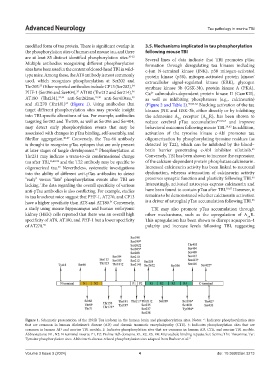
Figure 1. Schematic presentation of the 2N4R Tau isoform in the human brain and phosphorylation sites. Notes: *: Indicates phosphorylation sites
that are common in human Alzheimer’s disease (AD) and chronic traumatic encephalopathy (CTE). †: Indicates phosphorylation sites that are
common in human AD and murine TBI models, ‡: Indicates phosphorylation sites that are common in human AD, CTE, and murine TBI models.
Abbreviations: N1, N2: N-terminal inserts; P1, P2: Proline rich domains; R1, R2, R3, R4: Microtubule binding repeats; Ser: Serine; Thr: Threonine; Tyr:
Tyrosine phosphorylation sites. Alzheimer’s disease-related phosphorylation sites adapted from Basheer et al. 31
Volume 3 Issue 3 (2024) 4 doi: 10.36922/an.3213