
Page 146 - AN-3-4
P. 146
Advanced Neurology Drosophila Sirtuin 1 and Alzheimer’s disease
M
A B C D
E F G H
I J K L
N
O
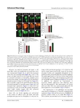
Figure 10. Fluorescence microscopy images and fluorescence intensity measurements showing anti-P-JNK, anti-Delta, and anti-NICD staining in the
third instar larval brain. (A-D) JNK expression, (E-H) Delta expression, and (I-L) NICD expression in the third instar larval brains of elav-Gal4/+;+/+;+/+,
elav-GAL4/+;+/+;UAS-ArcAβ /+, elav-Gal4/+;UAS-Sirt1/+;UAS-ArcAβ /+, and elav-Gal4/+;UAS-ArcAβ /+;UAS-Sirt1 RNAi /+. White arrowheads indicate
42
42
42
upregulated p-JNK, Delta, and NICD staining compared to elav-Gal4/+;+/+;+/+ (experimental control) flies (A, E, I). Scale bars indicate 10 μm (A-L).
(M-O) Histogram indicating average fluorescence intensity of P-JNK, Delta, and NICD in third instar larval brains of each genotype. P-JNK, Delta, and
NICD fluorescence intensities were analyzed using ImageJ software, NIH, USA. Error bars indicate mean ± standard error of the mean. Significance
was calculated using one-way analysis of variance with Tukey’s test in GraphPad Prism 5.0 and is indicated as ns: non-significant, *P < 0.05, **P <0.01,
***P < 0.0001. A total of 20 larval brains were observed for each genotype.
Abbreviation: NICD: Notch intracellular domain.
Moreover, we observed excessive cell death in AD status of JNK and Notch signaling in AD model flies. We
model flies, which was significantly decreased when Sirt1 noted a slight improvement in the rough eye phenotype,
was overexpressed (Figure 4E, H, and J) but increased abnormal bristles, and ommatidial arrangement in AD
when Sirt1 was downregulated (Figure 4F, I, and J). The model flies when Drosophila JNK (Bsk) was downregulated.
ectopic cell death observed in AD model flies was caused The pathological features of AD were further improved by
by increased expression of Drosophila apoptotic genes overexpressing Sirt1 along with downregulating Bsk in the
such as Grim, Reaper, and Hid (Figure 4K) and decreased/ AD model flies’ genetic background(Figure 5C, D, and E).
altered expression of diap1 in AD model flies (Figure 4L). In addition, we observed an improvement in
This was further supported by qRT-PCR analysis showing behavioral deficits (phototaxis and climbing) when
decreased expression of apoptotic genes (Figure 4K) and Bsk was downregulated. This improvement was further
increased diap1 expression (Figure 4L) when Sirt1 was enhanced when Sirt1 was overexpressed along with Bsk
overexpressed in the AD model flies’ genetic background. downregulation in AD model flies (Figure 5F and G).
These results further suggest that Sirt1 possesses The improvement in AD pathologies was associated
antiapoptotic properties (Figure 4E, H, J). with reduced cell death due to a decrease in apoptotic gene
Since JNK and Notch signaling are well-studied expression (Figure 5H) and the activation of inhibitors of
in the context of cellular stress, cell death, and early apoptotic proteins (IAPs) in AD model flies (Figure 5I).
developmental processes in Drosophila, we observed the This reduction was attributed to the decreased expression
Volume 3 Issue 4 (2024) 15 doi: 10.36922/an.4291