
Page 86 - AN-3-4
P. 86
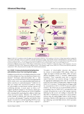
Advanced Neurology                                           SARS-CoV-2 in age-associated neurodegeneration


            Figure 4. SARS-CoV-2 persistence mimics the aging microenvironment in the brain. Viral persistence in the brain or distant organs induces an aging-like
            environment in the brain. SARS-CoV-2 infection accelerates the hallmarks of aging, which include sustained neuroinflammation, prolonged cytokine
            release, microglia activation, T-cell exhaustion, autoimmunity, deregulated UPR followed by plaque formation, increased oxidative stress, ER stress, and
            microbiome dysbiosis. All these events cumulatively promote neuronal loss and neurodegeneration. Image created with BioRender.com.
            Abbreviations: BBB: Blood–brain barrier; CD38: Cluster of differentiation 38; CTLA-4: Cytotoxic T lymphocyte antigen-4; ER: Endoplasmic reticulum;
            IL: Interleukin; Ki67: Kiel 67; PD1: Programmed cell death protein 1; ROS: Reactive oxygen species; SARS-CoV-2: Severe acute respiratory syndrome-
            coronavirus-2; TNF-α: Tumor necrosis factor alpha; UPR: Unfolded protein response.

            6.2. COVID-19-induced mitochondrial dysfunction    Disruption of mitochondrial dynamics also induces
            and oxidative stress: Fast-tracking brain aging    the release of mitochondrial DNA release, which can
            Oxidative stress and mitochondrial dysfunction play a central   trigger microglial activation and NOD-, LRR- and pyrin
            role in the pathogenesis of age-associated neurodegenerative   domain-containing protein 3 (NLRP3) inflammasome
                                                                       99
            diseases.   Prolonged  neuroinflammation  due  to  viral   formation.  These sequential events lead to persistent
                  95
            infection can increase the formation of reactive oxygen species   neuroinflammation  and  neuronal  damage.  In  addition,
            (ROS), which in turn cause mitochondrial dysfunction and   tau aggregation neurodegeneration may be caused by the
            further contribute to neuroinflammation.  SARS-CoV-2’s   activation of the NLRP3 inflammasome, triggered during
                                             96
                                                                                 27
            Nsps  can  interact with mitochondrial proteins, evading   SARS-CoV-2 infection.  Another hypothesis suggests that
            the mitochondria-mediated innate immune response and   the increased risk of AD in COVID-19 patients could be
                                                         97
            establishing infection. A  recent report by Duan  et al.    related to Aβ, which acts as an antimicrobial peptide. It
            suggests significant changes in mitochondria-related gene   is postulated that the SARS-CoV-2 neuroinvasion could
            expression and metabolic pathways in COVID-19 patients   promote Aβ generation as part of the immune response,
            after analyzing RNA sequencing data collected from lung   initiating the Aβ cascade and leading to extracellular Aβ
                                                                        100
            tissue and blood samples. Moreover, the SARS-CoV-2   deposition.  Furthermore, compared to control patients,
            protein ORF9b can directly alter mitochondrial function   patients with AD exhibit higher levels of ACE2 receptor
            to evade host cell immunity.  Together with ORF9b, ORF6   expression. Notably, ACE2 expression is not influenced by
                                  98
            impairs mitochondrial antiviral signaling (MAVS) protein   age, indicating a potential link between ACE2 expression
            function and suppresses the innate immune response.    and AD. 101
                                                         78

            Volume 3 Issue 4 (2024)                         13                               doi: 10.36922/an.4267