
Page 104 - AN-4-1
P. 104
Advanced Neurology TDP-43 regulates IFN1 production
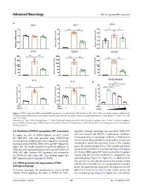
A
B
Figure 1. TDP43 suppresses TBK1-mediated IFN1 production. (A) Quantitative PCR analysis of IRF3, IRF7, IFN1, and related cytokine mRNA levels.
(B) IFN-β activity measured by dual-luciferase reporter assay. The data are shown as mean ± standard deviation (n = 5 per group). *P < 0.05, **P < 0.01,
and ***P < 0.001.
Abbreviations: TBK1: TANK-binding kinase 1; TDP43: TAR DNA-binding protein 43; IRF3: Interferon regulatory factor 3; IRF7: Interferon regulatory
factor 7; IFNA: Interferon-alpha; IFNB: Interferon-beta; ISG54: Interferon-stimulated gene 54; ISG56: Interferon-stimulated gene 56; IL6: Interleukin-6;
IL8: Interleukin-8.
3.4. Depletion of TDP43 upregulates IRF7 expression regulation through autophagy was examined. HEK-293T
To explore the effect of TDP43 depletion on IRF7, TDP43 cells were treated with MG132 (a proteasome inhibitor),
KO HEK-293T cells were generated using CRISPR-Cas9 chloroquine (CQ, an autophagy inhibitor), or Bafilomycin
technology. Immunoblot analysis was conducted to evaluate the A1 (A1, an autophagy inhibitor). Immunoblot analysis was
expression levels of TDP43, TBK1, IRF3, and IRF7 (Figure 4A, conducted to assess the expression levels of the targeted
Figure S4). The results revealed no significant difference in genes. The results revealed that p-TBK1 protein expression
TBK1 and IRF3 expression between wild-type and TDP43 KO was elevated in the MG132-treated group (Figure 5A, Figure
HEK-293T cells. However, IRF7 expression was increased in S5). Both p-TBK1 and p-IRF3 were increased in the groups
the TDP43-KO cells (Figure 4B). These findings suggest that treated with MG132+CQ or MG32+A1 compared to the
IRF7 expression may be regulated by TDP43. untreated group (Figure 5A, Figure S5). In addition, both
p62 and LC3-II were also increased in the groups treated
3.5. TDP43 promotes the degradation of TBK1 with MG132+CQ or MG132+A1 compared to the untreated
through autophagy group. No alteration was observed in the expression of p62
To further investigate the mechanism by which TDP43 and LC3-II between the MG132-only treated group and
inhibits IFN-β signaling, the effect of TDP43 on TBK1 the untreated group (Figure 5A, Figure S5). Consistently,
Volume 4 Issue 1 (2025) 98 doi: 10.36922/an.6272