
Page 109 - AN-4-4
P. 109
Advanced Neurology Incidental PFBC: Case report
tremors, and MRI of the brain showing extensive non- pathologic etiologies must be considered (Table 1). The
enhancing hyperintensities throughout the white matter of only physiologic or pathologic cause of brain calcifications
the frontal region (concerning for demyelinating disease) that manifests in a symmetrical fashion in both the basal
1,8
(Figure 2). Cerebrospinal fluid was negative for oligoclonal ganglia and dentate nuclei is PFBC, and mutational
bands, making multiple sclerosis an unlikely diagnosis. No analysis confirmed this diagnosis.
other potential etiologies for the white matter changes were Publications describing the various gene mutations
identified. An eventual CT scan of her brain, performed associated with PFBC and their phenotypes suggest that
due to the recent PFBC diagnosis in the father, showed subcortical white matter hyperintensities are not a common
calcifications in the basal ganglia (Figure 2). Genetic finding. However, there are rare reports of subcortical
testing on the daughter revealed the same heterozygous white matter changes in the brain in individuals with
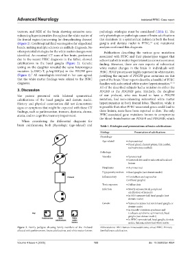
mutation (c.298C>T, p.Arg100Cys) in the PDGFB gene PFBC. PDGF proteins are highly expressed in white matter,
(Figure 3). All neurologists involved in her care agreed justifying the impacts of PDGFB gene mutations on this
7
that the white matter findings were related to the PFBC part of the brain. Four reports describe a handful of PFBC
9
diagnosis. families with subcortical white matter hyperintensities. 10-13
All of the described subjects had a mutation in either the
3. Discussion PDGFB or the PDGFRB gene. Similarly, the daughter
The patient presented with bilateral symmetrical of our proband, who was found to have a PDGFB
calcifications of the basal ganglia and dentate nuclei. mutation, had non-enhancing subcortical white matter
History and physical examination did not demonstrate hyperintensities in both frontal lobes. Therefore, while it
signs or symptoms that might be expected with these CT is possible that other PFBC-associated genes could lead to
findings, such as parkinsonism, tremors, dystonia, chorea, these lesions, none have been reported to date. The only
ataxia, and/or cognitive/memory impairment. PFBC-associated gene mutations known to compromise
the blood–brain barrier are PDGFB and PDGFRB, which
When considering the differential diagnosis for
brain calcifications, both physiologic (age-related) and Table 1. Etiologies and presentations of brain calcifications
Etiology Presentation of calcifications
Physiologic
Age-related • Asymmetrical
• Pineal gland, choroid plexus, falx cerebri,
and tentorium cerebelli
Pathologic
Vascular • Symmetrical
• Scattered dots and/or subcortical localized
lesions
Neoplasms • Asymmetrical
Hypoparathyroidism • Basal ganglia (not dentate nuclei)
Inflammatory • Cerebellum and suprasellar
(not basal ganglia)
Toxic exposure • Diffuse dots
Infection • Mostly asymmetrical, peripheral
calcification of lesion(s)
• In HIV: symmetrical, basal ganglia (not
dentate nuclei)
Genetic • Various locations but not in basal ganglia or
dentate nuclei
• In Aicardi–Goutières syndrome and
Cockayne syndrome: symmetrical, basal
ganglia (not dentate nuclei)
• In PFBC: symmetrical, basal ganglia, dentate
nuclei, thalami, subcortical white matter
Figure 3. Family pedigree showing family members of the Proband Abbreviations: HIV: Human immunodeficiency virus; PFBC: Primary
affected with parkinsonism, brain calcifications, and white matter lesions familial brain calcification.
Volume 4 Issue 4 (2025) 103 doi: 10.36922/an.4854