
Page 114 - AN-4-4
P. 114
Advanced Neurology CAA-related inflammatory case
A B
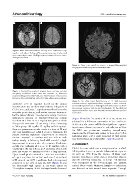
Figure 2. Fluid-attenuated inversion recovery shows asymmetrical high
signals in both sides of the brain. (A) A substantial amount of subcortical
white matter degeneration. (B) High signal intensity in the white matter
of the parietal cortex.
A B
Figure 4. There is no significant change in susceptibility-weighted
imaging microbleeds compared to before treatment.
A B
Figure 3. Susceptibility-weighted imaging shows extensive punctate
and patchy microbleeds in the cortex and subcortex. (A) Numerous
microhemorrhages were observable, and they were more pronounced in
the temporal lobe. (B) Multiple bilateral occipital lobe microhemorrhages.
Figure 5. The white matter hyperintensity on T2 fluid-attenuated
antibodies were all negative. Based on the clinical inversion recovery is significantly reduced compared to before treatment.
manifestations and auxiliary examinations, a diagnosis of (A) The high-signal intensity in the temporal lobe white matter has shown
improvement compared with the previous findings. (B) The abnormal
CAA-ri was considered. The patient was advised to receive signals in the cerebral cortex have decreased as compared to the previous
complete genetic testing and receive hormone treatment, examination.
but the patient’s family refused genetic testing. Therefore,
intravenous infusion of methylprednisolone sodium (Figure 5A and B). On January 23, 2024, the patient was
succinate at a dose of 1000 mg/day was given as shock admitted for a follow-up examination of the renal stent.
therapy (the dose was reduced every 3 days, decreasing At this time, the patient exhibited no significant cognitive
to 500 mg/day, 240 mg/day, 80 mg/day, and 40 mg/day); impairment and no recurrence of behavioral abnormalities.
then oral prednisone acetate tablets at a dose of 50 mg/ An MRI was performed, revealing demyelinating
day were administered. After 2 weeks of treatment, the changes on the T2 sequence similar to those observed in
patient exhibited significant improvement in memory September 2023, with a slight reduction in high signals
decline compared to baseline and was able to walk near the frontal and parietal cortex (Figure 6A and B).
independently. A follow-up head MRI revealed a slight
improvement in white matter degeneration. Prednisone 3. Discussion
acetate was continued at a dose of 45 mg/day, with a
weekly taper of 5 mg initiated post-discharge. One month CAA-ri is a rare autoimmune encephalopathy in which
later, the patient was readmitted due to a lung infection. Aβ deposition triggers a vascular inflammatory response.
Upon neurological examination, there was no evidence As early as 1979, from the biopsies of three CAA
of cognitive dysfunction or limb weakness. A repeat head patients’ brain tissues, some scholars found that amyloid
MRI showed that SWI microbleeds had not progressed deposits (showing orange-red in Congo red staining)
compared to the MRI on July 18, 2024 (Figure 4), and were widespread in the leptomeningeal and cerebral
the abnormally high signals in the white matter on cortical blood vessels. Moreover, lymphocyte infiltration
T2-weighted imaging FLAIR had significantly decreased was detected in and around these deposits. Through
3
Volume 4 Issue 4 (2025) 108 doi: 10.36922/AN025080015