
Page 87 - AN-4-4
P. 87
Advanced Neurology Cytokine response to EV therapy in SCI
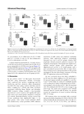
Figure 3. Comparison of cytokine levels. Boxplots display the concentrations of G-CSF, IL-10, IL-1β, IL-5, IL-17A, and RANTES across four experimental
groups: SCI (untreated), SCI FM (treated with FM only), SCI FM+EVs5 (treated with 5 µg EVs in FM), and SCI FM+EVs10 (treated with 10 µg EVs in
FM). *p<0.05, ANOVA with Tukey’s post hoc test.
Abbreviations: G-CSF: Granulocyte colony-stimulating factor; EVs: Extracellular vesicles; FM: Fibrin matrix; IL: Interleukin; SCI: Spinal cord injury.
the concentrations of pro-inflammatory IL-1β (1.5-fold) modulation of both central and peripheral conduction
and IL-5 (1.9-fold), as well as the anti-inflammatory pathways. MEP registration frequency progressively
IL-10 (2.4-fold) relative to SCI FM. decreased over time in all SCI groups, whereas SSEP
A similar trend was observed for IL-17A levels, where a amplitudes showed preserved peripheral responses but
1.6-fold decrease (p<0.05) was found in the SCI FM+EVs10 impaired cortical signals. Histological and morphometric
analyses revealed greater tissue preservation and
group compared to the SCI FM control group (Figure 3E).
Notably, RANTES expression (Figure 3F) was significantly oligodendrocyte survival in EV-treated groups, with a
higher in the SCI FM+EVs5 group than in the SCI group more pronounced neuroprotective effect at higher doses.
These findings provide a strong foundation for the present
(4-fold, p<0.05). In addition, RANTES levels were 2-fold
higher in SCI FM compared with the SCI group (p<0.05). investigation into the inflammatory responses following
MSC-EV application in the same SCI model.
4. Discussion We have previously shown that MSCs embedded in
12
In our previous study, we thoroughly characterized an FM can retain their viability at the SCI site for up to
MSC-EVs using transmission electron microscopy, 60 days and migrate rostrally and caudally for more than
16
which confirmed their uniform morphology and size 5 mm. In this study, we conducted a multiplex analysis
distribution, and flow cytometry, which demonstrated of 23 cytokines in rat spinal cord homogenates at 60 dpi—
that MSC-EVs share surface markers with their parental representing the chronic phase of SCI—after the treatment
MSCs, including Sca-1, CD49e, and CD44. Functional with FM combined with MSC-EVs at two dosages (5 µg
assessments, including Basso, Beattie, and Bresnahan and 10 µg). The results were compared to SCI without
(BBB) locomotor testing, revealed a dose-dependent treatment and SCI treated with FM alone. Our analysis
improvement in motor recovery, with significantly higher revealed an upregulation of several cytokines following
BBB scores in EV-treated groups compared to untreated MSC-EV therapy.
SCI controls. Electrophysiological analysis showed that FM treatment alone was associated with a sustained
M-wave amplitudes were significantly higher in the increase of multiple chemokines (G-CSF, M-CSF, GM-CSF)
same groups, while motor evoked potentials (MEP) and predominantly pro-inflammatory effects (IL-1β, IL-2,
and somatosensory evoked potentials (SSEP) indicated IL-5, IL-6, IL-7, IL-17A, IL-18, IFN-γ, and MIP-3α),
Volume 4 Issue 4 (2025) 81 doi: 10.36922/AN025110022