
Page 16 - ARNM-1-1
P. 16
Advances in Radiotherapy
& Nuclear Medicine Seed implantation head-and-neck carcinoma
region. The recurrence site location varies, resulting in
differences in anesthesia style, set-up position, and template
model.
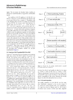
The workflow of 3D-PT guidance I-125 RSI-BT is as
follows: (i) Patients are immobilized through fixation with
vacuum pad followed by a contrast CT simulation with
5 mm thickness slices; (ii) the CT images are transferred
into the BT-TPS, the doctors define and delineate the
targets, OAR, the CTV=GTV + 5 − 6 mm, the PD is
110 – 130 Gy for squamous cell carcinoma and 130 –
160 Gy for soft-tissue carcinoma, and the activity of I-125
seed was 0.4 – 0.6 mCi; (iii) needle pathway is designed
to optimally satisfy the best conformality, a 3D-printer is
utilized to print the individual template; (iv) the patients
are again set up and the 3D-PNCT is fixed to the patient’s
body or the 3D-PCT is fixed with a stabilization frame
connected to the CT couch; (v) a CT scan is performed to
visualize the endpoint of each of the needles position; (vi)
a fine adjustment is made if the needle position error more
than 2 mm; (vii) I-125 seeds are implanted according to the
pre-plan; and (viii) the CT scan is again performed and the
slices transferred to the BT-TPS for post-plan dosimetry
evaluation . These steps must be strictly followed for
[40]
quality control throughout RSI-BT procedures to ensure
the post-plan dose evaluation satisfies the pre-plan
requirements (Figure 2).
5.5. Toxicity of 3D-PT-guided RSI-BT for HNC
Jiang et al. reported a study on 42 patients with recurrent
[17]
HNC who received 3D-PNCT-assisted CT-guided I-125
RSI-BT. The activity of the seeds was 0.34 – 0.7 mCi
(median: 0.6); the number of seeds implanted was 10 – 126
(median: 34); the number of needles implanted was 4 – 31
(median: 11); and the mean D2cc (dose to the most exposed
2 cc volume) and D0.1cc (dose to the most exposed 0.1 cc
volume) of the skin was 24.9 Gy (7.1 – 85.5) and 47.5 Gy
(9.4 – 167.2), respectively, whereas those of the spinal Figure 2. Protocol for 3D printing template-assisted computed
cord was 8.4 Gy (4.5 – 33.3) and 14.2 Gy (13.6 – 63.0), the tomography-guided seed implantation.
mucosa was 35.1 Gy (4.2 – 82.8) and 87.0 Gy (6.6 – 214.1),
parotid glands were 16.2 Gy (12.8 – 19.7) and 29.8 Gy between the affected and unaffected sides for the ramus
(26.1 – 33.4), and the trachea was 17.9 Gy (2.5 – 45.9) (p = 0.005; p < 0.05) and the body (p = 0.043; p < 0.05).
and 32.7 Gy (3.9 – 83.9), respectively. No case had an Pediatric parotid gland carcinoma survivors treated with
acute reaction to Grade ≥3. One case had a Grade 3 nerve interstitial BT experienced mild effects on the mandibular
response. Wu et al. observed significant differences in growth, particularly in the ramus. However, the overall
[43]
3D cephalometry measurements between the affected and impact was relatively minimal in these survivors.
unaffected sides of the mandible. The median fore-and-aft 5.6. I-125 seed migration
increments of the condyle length, the ramus length, and
the mandibular body length were 1.23 mm, 0.19 mm, and The quality assurance of I-125 RSI-BT was the target
1.66 mm, respectively, for the affected side. In contrast, dose distribution compared to the pre-plan design. If
these measurements were 1.37 mm, 1.95 mm, and the radioactive seeds migrated after seed implantation,
3.42 mm, respectively, for the unaffected side. Statistical the target dose may be reduced, and the risk of tumor
analysis revealed a significant difference in increments recurrence may increase. At the same time, migrated
Volume 1 Issue 1 (2023) 8 https://doi.org/10.36922/arnm.0907