
Page 18 - ARNM-2-1
P. 18
Advances in Radiotherapy
& Nuclear Medicine Blood glucose and I-123 MIBG lung uptake
Figure 3. Mean 24-h lung-to-mediastinum ratio (LMR) in diabetic and
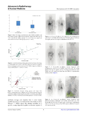
non-diabetic groups. Diabetic group showed a mean 24-h LMR value of Figure 6. A 67-year-old diabetic man diagnosed with paraganglioma
1.38 ± 0.20 and non-diabetic group 1.17 ± 0.14. There was a significant underwent I-123 MIBG whole-body scan. His fasting glucose level was
mean difference between these groups (0.21; P < 0.001). 236 mg/dL, and his 24-h lung-to-mediastinum ratio was 1.73.
Figure 4. Correlation between fasting glucose level and mean 24-h lung-
to-mediastinum ratio (LMR). There was a significant positive correlation
between fasting glucose level and mean 24-h LMR, irrespective of diabetic
status, with Pearson’s r = 0.596 (P < 0.001). Figure 7. A 63-year-old non-diabetic woman diagnosed with
paraganglioma underwent I-123 MIBG whole-body scan. Her fasting
glucose level was 93 mg/dL, and her 24-h lung-to-mediastinum ratio
(LMR) was 1.05, which was lower than the LMR of a representative
diabetic patient (Figure 6).
Figure 5. Correlation between fasting glucose and mean 24-h
lung-to-mediastinum ratio in diabetic and non-diabetic groups. Pearson’s
correlation analysis revealed significant positive correlations, separately, in
diabetic (r = 0.397; P = 0.016) and non-diabetic groups (r = 0.579; P < 0.001).
synthesis, storage, and reuptake, but in some tissues, Figure 8. An 83-year-old non-diabetic woman diagnosed with
peptide hormone or neuromodulator is the main secretory pheochromocytoma underwent I-123 MIBG whole-body scan. Her
fasting glucose level was 107 mg/dL with a 24-h lung-to-mediastinum
element. 1-3,19 MIBG enters the adrenal medullae by a ratio of 1.43. The scan indicates that MIBG uptake could also increase in
specific, energy-dependent uptake mechanism (uptake 1) non-diabetic patients.
Volume 2 Issue 1 (2024) 4 https://doi.org/10.36922/arnm.2506