
Page 43 - ARNM-2-1
P. 43
Advances in Radiotherapy
& Nuclear Medicine Exploring F-FDG PET/CT in Richter transformation
18
male predominance (i.e., eight male [66%] and four female marrow, or lymph nodes. CLL is considered an indolent
5
[34%] patients) among the patients. From the F-FDG or low-grade lymphoma, and the CLLs of 5%–10% of
18
PET/CT scans, eight (66%) scans were positive, and patients reportedly develop into an aggressive form
four (34%) scans were negative. Patients with a positive through RT. 6-10
PET/CT result subsequently underwent histopathological The use of F-FDG PET/CT can effectively identify
18
examination. Among them, three individuals had evidence biopsy sites in tumors (i.e., intense metabolically active
of RT, particularly in the form of DLBCL. nodes), and in combination with other biological
The patients started treatment for CLL before markers, physicians can accurately assess the CLL stage
confirming RT, and the treatment included active and prognosis of patients to make better, more informed
monitoring, chemotherapy, and immunotherapy. treatment decisions. 7,8
Furthermore, the three RT-positive patients adopted the CLL is characterized by low F-FDG avidity in PET/CT
18
R-CHOP chemotherapy regimen. In general, most CLL imaging, corresponding to the low mitotic activity of the
patients can lead a prolonged life with the disease, while lymphocytes. However, RT would increase the avidity
3-9
some CLL patients may have quicker CLL progression. of lymphoid tissues to F-FDG (Figure 1). The increased
18
Unfortunately, three patients with untransformed CLL SUVmax in lymph nodes correlated with high suspicion for
were deceased during the study. RT and should be further evaluated. Furthermore, F-FDG
18
The specificity of F-FDG PET/CT for detecting RT was PET/CT may be used to identify intensely metabolically
18
44.5%, with positive and negative predictive values of 37.5% active nodes for biopsy. 10
and 100%, respectively. Frequencies of cases with increased The main value of F-FDG PET/CT in CLL patients is
18
uptake in the “only nodal sites” and “nodal and extranodal its ability to detect RT with a negative predictive value of
sites,” relative to positive biopsies, were 20% and 66%, 97% and identify sites of increased F-FDG uptake that is
18
respectively. Cervical (28.5%) and inguinal (28.5%) lymph suitable for biopsy or surveillance. In our study, cervical
3
nodes were the most affected in the supradiaphragmatic and inguinal lymph nodes were, respectively, the most
and infradiaphragmatic areas, respectively. The complete affected in the supradiaphragmatic and infradiaphragmatic
18 F-FDG PET/CT results are displayed in Tables 1-3. areas, respectively (Figures 1 and 2). The SUVmax in the
4. Discussion cervical nodes had a range of 6.1–7.7, but the SUVmax was
more intense in the inguinal lymph nodes with a range of
CLL is defined by the proliferation of phenotypically 6.8–26.1. Our findings also displayed that increased uptake
monoclonal B-cell lymphocytes derived from blood, in the nodal and the extranodal sites were predictive of
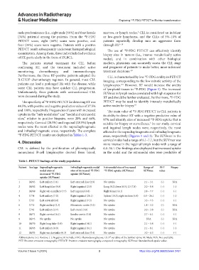
Table 1. PET/CT findings of the study population
Patient Sex/age Supradiaphragmatic Infradiaphragmatic nodal Extranodal sites of increased Range of PET Biopsy
nodal sites of sites of increased F‑FDG 18 F‑FDG uptake (SUVmax) SUVmax value
18
increased F‑FDG uptake (SUVmax)
18
uptake (SUVmax)
1 M/63 Left axillary (3.1) Left external iliac (2.9) No uptake 2.1 – 3.1 (-) N/A
2 M/62 Left lung hilar (8.8) Right inguinal (2.0) Lung (8.2); liver (6.5); L3 (7.8) 2.0 – 8.8 (+) (+)
3 M/68 Right sub maxillary (7.7) Left inguinal (6.8) Right femur (4.1) 2.8 – 7.7 (+) (-)
4 F/74 Left axillary (7.0) Right inguinal (26.1) Spleen (10.3); right ischion (6.4) 1.8 – 26.1 (+) (+)
5 F/53 Left cervical (6.6) Right inguinal (7.5) No uptake 3.5 – 7.5 (+) (-)
6 F/71 Right axillary (2.3) Mesenteric nodes (3.2) No uptake 1.8 – 3.2 (-) N/A
7 F/76 Left axillary (2.7) Left crural (3.8) No uptake 2.0 – 3.8 (-) N/A
8 M/71 Right cervical (6.1) Lombo-aortic (3.8) No uptake 2.7 – 6.1 (+) (-)
9 M/73 No uptake No uptake No uptake N/A (-) N/A
10 M/70 Right lung hilar (5.8) Right inguinal (4.1) No uptake 2.1 – 5.8 (+) (-)
11 M/65 Left axillary (8.0) Right inguinal (8.1) No uptake 2.1 – 8.1 (+) (+)
12 M/53 Right sus clavicular (6.3) Left external iliac (5.4) No uptake 3.0 – 6.3 (+) (-)
Abbreviations: (+): Positive; (-): Negative; F: Female; FDG: Fluorodeoxyglucose; L3: 3 vertebra of the lumbar spine; M: Male; N/A: Not available;
rd
PET: Positron emission tomography; PET/CT: Positron emission tomography-computed tomography; SUVmax: Standardized uptake value.
Volume 2 Issue 1 (2024) 3 https://doi.org/10.36922/arnm.2431