
Page 44 - ARNM-2-1
P. 44
Advances in Radiotherapy
& Nuclear Medicine Exploring F-FDG PET/CT in Richter transformation
18
Table 2. Percentage of increased F‑FDG uptake in lymph Table 4. Summary of PET/CT predictive values for detecting
18
node areas RT nodes % of increased uptake
Lymph nodes Percentage of increased F‑FDG uptake (%) Study RT SUVmax Specificity Predictive value (%)
18
Cervical 28.5 cases (%) Positive Negative
Inguinal 28.5 Bruzzi et al. 3 10 5 80 53 97
Axillary 14 Mauro et al. 4 17 5 71.2 51.3 94
Lung hilar 14 Falchi et al. 12 95 5 47 38 92
Lombo-aortic 14 Abbreviations: PET/CT: Positron emission tomography-computed
Other 1 tomography; RT: Richter transformation; SUVmax: Standardized
uptake value.
Abbreviation: FDG: Fluorodeoxyglucose.
A B
Table 3. Predictive value of PET/CT for detecting RT in 12
patients
Parameters Value
PET value (number of patients)
Positive 4
Negative 8
Predictive values (%)
Positive 37.5
Negative 100
Specificity (%) 44.5
False positive (%) 62.5
Nodal sites (%)
Nodal and extranodal sites 37.5
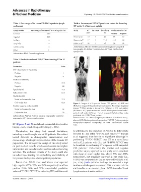
Only nodal sites 62.5 Figure 1. Images of a 53-year-old female CLL patient. (A) MIP and
Positive biopsy at nodal sites (%) (B) fusion images of the patient’s coronal section. The images displayed
intense F-FDG uptake in the cervical (SUVmax = 6.5) and axillary
18
Nodal and extranodal sites 66
(SUVmax = 6.3) lymph nodes and low F-FDG uptake in the lombo-
18
Only nodal sites 20 aortic lymph nodes (SUVmax = 4.3). A biopsy of the left axillary nodes
Abbreviations: PET/CT: Positron emission tomography-computed performed after PET/CT was positive.
tomography; RT: Richter transformation. Abbreviations: CLL: Chronic lymphocytic leukemia; FDG: Fluoro-deoxy-
glucose; MIP: Maximum intensity projection; PET/CT: Positron emission
tomography-computed tomography; SUVmax: Standardized uptake
RT (Figures 2 and 3) (nodal and extranodal sites/positive value.
biopsy = 66%; nodal sites/positive biopsy = 20%).
Nonetheless, the study had several limitations, be attributed to the limitation of PET/CT to differentiate
18
including a small sample size of 12 patients. The cohort between RT and other F-FDG-avid tumors. 3,4,12 Papajik
lacked diversity in demographic characteristics and et al. suggested that there is no significant advantage in
18
disease stage, affecting its representation of the broader RT performing F-FDG-PET/CT over CT as a surveillance
18
population. The retrospective design of this study relied tool in CLL patients with CLL, but F-FDG-PET/CT may
on past medical records, which could contain incomplete be beneficial in confirming RT diagnosis in RT-suspected
11
information and introduce selection bias and confounding CLL patients. Collectively, the positive and negative
variables. The exclusion criteria of the study could also predictive values reported in our study were consistent
introduce bias, limiting the applicability and reliability of with those of other studies (i.e., above 37% and 100%,
the present findings to a diverse patient population. respectively), but specificity reported in our study (i.e.,
44.5%) was comparable to that as reported by Falchi et al.
Bruzzi et al., Falchi et al., and Mauro et al. reported (i.e., 47%) (Table 4). Our findings also revealed a high
12
high negative (97%, 92%, and 94%, respectively) and low incidence of false positive cases (62.5%), and this could be
positive (53%, 38%, and 51%, respectively) predictive attributed to an accelerated phase of CLL instead of frank
values of F-FDG PET/CT for detecting RST, and this could lymphomatous transformation. 4
18
Volume 2 Issue 1 (2024) 4 https://doi.org/10.36922/arnm.2431