
Page 45 - ARNM-2-1
P. 45
Advances in Radiotherapy
& Nuclear Medicine Exploring F-FDG PET/CT in Richter transformation
18
A B acknowledge the limitations identified in the study. The
retrospective nature, small sample size, and potential
bias in patient selection challenged the reliability and
practicality of the findings. Despite these constraints, the
study highlighted the significance of PET/CT imaging
in confirming clinical suspicions and guiding biopsy site
selection. Moving forward, larger prospective studies with
diverse cohorts are warranted to enhance the robustness of
C evidence and provide a more comprehensive understanding
of the role of PET/CT scans in RT evaluation.
Acknowledgments
None.
Funding
None.
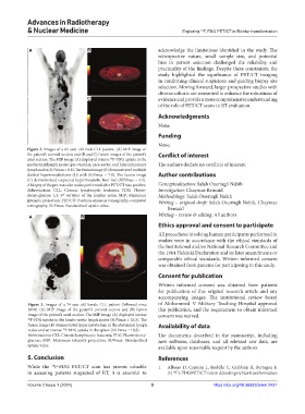
Figure 2. Images of a 62-year-old male CLL patient. (A) MIP image of
the patient’s coronal section and (B and C) fusion images of the patient’s Conflict of interest
18
axial section. The MIP image (A) displayed intense F-FDG uptake in the
mediastinal lymph nodes (pre-vascular, para-aortic, and hilar pulmonary The authors declare no conflicts of interest.
lymph nodes; SUVmax = 8.8). The fusion image (B) demonstrated multiple
skeletal hypermetabolisms (L3 with SUVmax = 7.8). The fusion image Author contributions
(C) demonstrated suspected hypermetabolic liver foci (SUVmax = 6.5).
A biopsy of the pre-vascular nodes performed after PET/CT was positive. Conceptualization: Salah Oueriagli Nabih
Abbreviations: CLL: Chronic lymphocytic leukemia; FDG: Fluoro- Investigation: Chaymae Bensaid
rd
deoxy-glucose; L3: 3 vertebra of the lumbar spine; MIP: Maximum Methodology: Salah Oueriagli Nabih
intensity projection; PET/CT: Positron emission tomography-computed Writing – original draft: Salah Oueriagli Nabih, Chaymae
tomography; SUVmax: Standardized uptake value.
Bensaid
A B Writing – review & editing: All authors
Ethics approval and consent to participate
All procedures involving human participants performed in
studies were in accordance with the ethical standards of
the Institutional and/or National Research Committee and
the 1964 Helsinki Declaration and its later amendments or
comparable ethical standards. Written informed consent
was obtained from patients for participating in this study.
Consent for publication
Written informed consent was obtained from patients
for publication of this original research article and any
accompanying images. The institutional review board
Figure 3. Images of a 74-year-old female CLL patient (followed since of Mohammed V Military Teaching Hospital approved
2018). (A) MIP image of the patient’s coronal section and (B) fusion this publication, and the requirement to obtain informed
image of the patient’s axial section. The MIP image (A) displayed intense consent was waived.
18 F-FDG uptake in the lombo-aortic lymph nodes (SUVmax = 26.3). The
fusion image (B) demonstrated hypermetabolism in the abdominal lymph Availability of data
nodes and an intense F-FDG uptake in the spleen (SUVmax = 10.3).
18
Abbreviations: CLL: Chronic lymphocytic leukemia; FDG: Fluoro-deoxy- The documents described in the manuscript, including
glucose; MIP: Maximum intensity projection; SUVmax: Standardized new software, databases, and all relevant raw data, are
uptake value. available upon reasonable request by the authors.
5. Conclusion References
While the F-FDG PET/CT scan has proven valuable 1. Albano D, Camoni L, Rodella C, Giubbini R, Bertagna F.
18
in assessing patients suspected of RT, it is essential to 2-[ F]-FDG PET/CT role in detecting richter transformation
18
Volume 2 Issue 1 (2024) 5 https://doi.org/10.36922/arnm.2431