
Page 53 - ARNM-2-1
P. 53
Advances in Radiotherapy
& Nuclear Medicine Bone scan image features of secondary syphilis
features of osteolytic lesions caused by secondary syphilis whole right knee joint and proximal tibia, indicating
in the past decades. a rare inflammatory bone lesion beyond the facture
We herein present the case of a 68-year-old woman (Figure 1A and C). The inflammatory nature of the lesion
complaining of pain of the right knee, which was initially prompted us to query the patient again. Further disclosure
misdiagnosed by plain radiography as fracture on inner from her revealed certain vital diagnostic clues: She had
right tibia plateau but was finally clarified as osteolytic been diagnosed with syphilis, acquired from her husband
lesion due to secondary syphilis affecting the whole right through an intimate infection, and had been cured after
knee. undergoing a penicillin treatment for a month 8 years
ago. Based on the positive results from non-treponemal
2. Case presentation test (TRUST 1:4) and Treponema pallidum antibody test
(403.54 s/co), with the latter registering an increased
A 68-year-old housewife without any significant medical antibody titer in serum, we ruled that the patient was
history was admitted to the hospital for the pain afflicted diagnosed with osteolytic lesion caused by secondary
to her right knee, a condition that lasted for 10 days before syphilis, which was validated by Warthin–Starry silver
admission and exhibited poor response to painkiller. staining test (Figure 1F). Accordingly, the patient was
A radiological examination initially indicated to the prescribed a course of antibiotics treatment and also
patient revealed a serious osteolytic lesion on inner right surgically treated with total knee arthroplasty (Figure 1G).
tibia plateau (Figure 1B and D), which was suggestive of
obsolete fracture, but the patient denied having suffered 3. Discussion
from knee injury. Hematoxylin and eosin staining, on the Single-photon emission computed tomography (SPECT)
other hand, showcased significant lymphocyte infiltration scans using radiotracers for bone such as 99m Tc-methylene
in the center of the lesion but histopathologic features diphosphonate ( Tc-MDP) could provide detailed
99m
that could reveal the cause of osteolytic lesion were not information about the physiological and anatomical state
observed (Figure 1E). Without knowing the exact cause of bone, which could help with the detection of early
of the fracture rendered the administration of appropriate bony lesions of secondary syphilis. Imaging bone with
clinical treatment for the patient impossible. 99m Tc-MDP SPECT remains the mainstay of bone lesion
Therefore, a bone scanning examination was diagnosis, despite the rapid advances in other technologies,
performed. The imaging results showed intense uptake such as positron emission tomography (PET/CT) and
of 99m Tc-methylene diphosphonate ( Tc-MDP) in the magnetic resonance imaging (MRI).
99m
A B C D
E F G
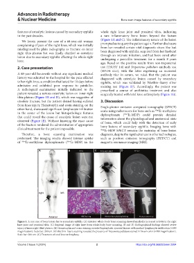
Figure 1. A rare case of bone lesion due to secondary syphilis. (A) Anterior whole-body bone scanning showed markedly increased activity in the right
knee joint and proximal tibia. (C) Regional image of right knee from whole-body bone scanning. (B and D) Radiographical findings showed severe
injury of inner right tibial plateau. (E) Hematoxylin and eosin staining revealed hyperplastic synovial tissues with marked lymphocytic infiltration (×100
magnification). Scale bar: 200 um. (F) Warthin–Starry staining revealed the presence of Treponema pallidum stained in brown color (×100 magnification).
Scale bar: 200 um. (G) Treatment of total knee arthroplasty.
Volume 2 Issue 1 (2024) 2 https://doi.org/10.36922/arnm.2204