
Page 77 - ARNM-2-2
P. 77
Advances in Radiotherapy
& Nuclear Medicine Wound reconstruction with Intergra®
A B C D E F
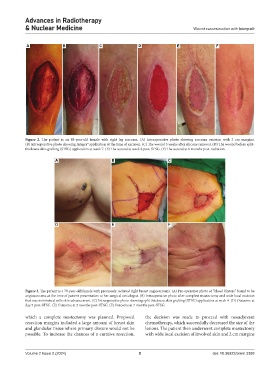
Figure 2. The patient is an 88-year-old female with right leg sarcoma. (A) Intraoperative photo showing sarcoma excision with 2 cm margins.
(B) Intraoperative photo showing Integra® application at the time of excision. (C) The wound 5 weeks after silicone removal. (D) The wound before split-
thickness skin grafting (STSG) application at week 7. (E) The wound at week 8 post-STSG. (F) The wound at 6 months post-radiation.
A B C
D E F
Figure 3. The patient is a 79-year-old female with previously radiated right breast angiosarcoma. (A) Pre-operative photo of “blood blisters” found to be
angiosarcoma at the time of patient presentation to her surgical oncologist. (B) Intraoperative photo after complete mastectomy and wide local excision
that was minimized with skin advancement. (C) Intraoperative photo showing split thickness skin grafting (STSG) application at week 4. (D) Outcome at
day 5 post-STSG. (E) Outcome at 2 months post-STSG. (F) Outcome at 7 months post-STSG.
which a complete mastectomy was planned. Proposed the decision was made to proceed with neoadjuvant
resection margins included a large amount of breast skin chemotherapy, which successfully decreased the size of the
and glandular tissue where primary closure would not be lesions. The patient then underwent complete mastectomy
possible. To increase the chances of a curative resection, with wide local excision of involved skin and 2 cm margins
Volume 2 Issue 2 (2024) 5 doi: 10.36922/arnm.3388