
Page 74 - ARNM-2-2
P. 74
Advances in Radiotherapy
& Nuclear Medicine Wound reconstruction with Intergra®
proliferative capacity of surviving cells, decreased Herein, we present a case series of four patients who
vascularity, vessel thrombosis, inhibition of collagen were successfully treated after cancer excision with
synthesis, both acute and chronic inflammation, and Integra® and skin grafting followed by adjuvant radiation,
ultimately obliterative endarteritis and tissue necrosis. 12-21 as well as two patients treated in the same manner but in
the setting of a previously radiated area for prior cancer
The challenging nature of reconstructing irradiated
wounds has led to novel ideas of wound management. For treatment (Table 1). A review of the literature on the utility
of RDMs to improve reconstructive outcomes in radiated
example, Mehanni et al. investigated the use of stem cells fields was performed to better understand its emerging role
to heal irradiated wounds. More commonly, surgeons to manage these complex cases. Finally, by comparing our
22
are turning to engineered regenerative dermal matrices experience with the systematic review of the literature, we
(RDMs) to heal irradiated wounds. Animal-derived RDMs describe our algorithm for approaching reconstructions
have populated the market, including but not limited to using RDMs when radiation is involved.
porcine (PriMatrix®), piscine (Kerecis), ovine (OviTex),
and bovine (Integra®) derivatives. 2. Methods and materials
Integra® (Integra Life Sciences, Plainsboro, NJ) is the We performed a case review of patients successfully treated
first such product to be commercially available, initially after large cutaneous cancer excision with our algorithm
designed in the 1970s and approved for use in burn for staged wound reconstruction using RDM followed by
reconstruction in 1996. The utility of RDMs has since skin thickness skin grafting. Our algorithm began with
23
seen wide expansion to include a variety of different placement of RDM at the time of cancer excision even in
uses and applications. 24-41 The use of dermal regenerative the presence of exposed vital structures such as bone or
templates in conjunction with skin grafting outside the tendon. This constituted the first stage of reconstruction.
setting of radiation is well represented in the literature. 42-47 Once negative margins were assured, the Integra® was left
By supporting the creation of a viable recipient wound in place for 3 – 4 weeks, after which time the silicone layer
bed, RDMs can expand graft reconstruction options after was removed in the clinic. The matrix was left in place for
a variety of surgically created wounds. 27,28,48-50 This includes an additional 1 – 2 weeks, to allow granulation tissue to
coverage of vital structures after surgical excision of reach surface level and resolve any contour irregularities.
tumors, a reconstruction previously thought to require a Coverage of vital structures was then assessed, and if
more complex flap type procedure. 51 successful, split-thickness skin grafting was performed.
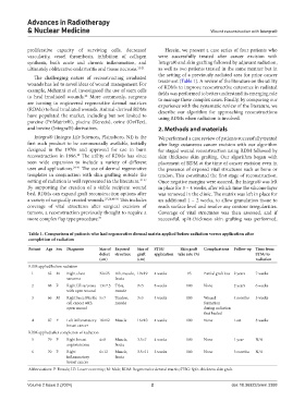
Table 1. Comparison of patients who had regenerative dermal matrix applied before radiation versus application after
completion of radiation
Patient Age Sex Diagnosis Size of Exposed Size of STSG Skin graft Complications Follow‑up Time from
defect structure graft application take rate (%) STSG to
(cm) (cm) radiation
RDM applied before radiation
1 62 M Right chest 32×25 Rib, muscle, 19×19 4 weeks 95 Partial graft loss 2 years 7 weeks
sarcoma fascia
2 88 F Right LE sarcoma 13×7.5 Tibia, 9×5 8 weeks 100 None 2 years 6 weeks
with open wound muscle
3 66 M Right knee Merkle 5×7 Tendon, 3×5 4 weeks 100 Wound 4 months 5 weeks
cell cancer with muscle formation
open wound during radiation
that healed
4 87 F Left inflammatory 18×12 Muscle 16×10 4 weeks 100 None Lost 8 weeks
breast cancer
RDM applied after completion of radiation
5 79 F Right breast 4×8 Muscle, 3.5×7 4 weeks 100 None 1 year N/A
angiosarcoma fascia
6 70 F Right 6×12 Muscle, 3.5×11 5 weeks 100 None 3 months N/A
inflammatory fascia
breast cancer
Abbreviations: F: Female; LE: Lower extremity; M: Male; RDM: Regenerative dermal matrix; STSG: Split-thickness skin graft.
Volume 2 Issue 2 (2024) 2 doi: 10.36922/arnm.3388