
Page 70 - ARNM-2-2
P. 70
Advances in Radiotherapy
& Nuclear Medicine 18 F-FDG PET/CT in LVAD infection
A
B
C
D
E
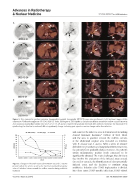
Figure 2. Five consecutive positron emission tomography/computed tomography (PET/CT) scans were performed. (A-E) Sectional images of five
consecutive F-fluorodeoxyglucose ( F-FDG) PET/CT scans. The degree of FDG uptake in the infection lesions around the outflow cannula (arrow in
18
18
A-E; maximum standardized uptake value was 9.4, 5.7, 3.7, 2.9, and 2.5, respectively) gradually decreased after antibiotic treatment. The abnormal FDG
uptake along the driveline (arrowhead) did not significantly change, indicating post-operative reactive changes rather than infection.
and extent of the infective areas is instrumental in making
clinical treatment decisions. Culture of both blood
9
and the area in question around the outflow cannula
at the abdominal surgical area indicated an infection
with E. cloacae and S. aureus. After a series of invasive
debridement procedures and targeted antibiotic treatment,
the patient’s fever gradually abated. However, the patient’s
serum inflammatory marker levels continued to be
significantly elevated. The main challenges that clinicians
face involve the evaluation of the infected areas around
the outflow cannula, the identification of other potentially
Figure 3. Changes in the erythrocyte sedimentation rate and C-reactive infected areas, and the decision to continue using
protein levels in the serum, as well as the maximum standardized uptake
value of infection lesions around the outflow cannula during antibiotic antibiotics. Infection after LVAD implantation is divided
treatment into three types: LVAD-specific infections, LVAD-related
Volume 2 Issue 2 (2024) 3 doi: 10.36922/arnm.3690