
Page 69 - ARNM-2-2
P. 69
Advances in Radiotherapy
& Nuclear Medicine 18 F-FDG PET/CT in LVAD infection
( F-FDG PET/CT). FDG PET/CT has an incremental patient’s body temperature gradually returned to normal.
4-7
18
role in technically limited or inconclusive cases of computed However, the levels of inflammatory markers, such as
tomography (CT) or echocardiography. Occasionally, it the erythrocyte sedimentation rate (ESR) and C-reactive
8
is difficult to distinguish high fluorodeoxyglucose (FDG) protein (CRP) levels, remained abnormal. The patient was
uptake lesions caused by infection and those resulting referred to the nuclear medicine department for PET/CT
from post-operative changes. The combination of PET/CT examination to investigate the cause. The first PET/CT scan
and serum inflammatory markers can help improve the displayed abnormally increased FDG uptake around the
diagnosis and evaluation of infected areas. outflow cannula (oval in Figure 1), which could indicate
infected foci or reactive changes after debridement surgery.
2. Case presentation The radioactivity at the post-operative sternum site (black
A 60-year-old man who presented with fever and elevated arrowhead in Figure 1) and along the driveline may represent
serum inflammatory marker levels after LVAD implantation aseptic inflammatory reactions. Based on the above PET/CT
presented to our hospital for treatment. The patient was results, the patient continued taking levofloxacin orally, and
diagnosed with myocarditis at the age of 17 with dilated the serum inflammatory markers were regularly monitored.
cardiomyopathy at the age of 50. Finally, the patient The following year, the patient underwent five consecutive
underwent LVAD implantation surgery as destination PET/CT scans to evaluate his condition (Figures 1 and 2).
therapy due to end-stage heart failure at the age of 59. The The abnormally increased FDG uptake in areas around
patient developed a fever after 49 days of LVAD implantation. the outflow cannula (oval in Figure 1; arrows in Figure 2)
Blood cultures indicated an infection with Enterobacter gradually disappeared after antibiotic treatment. However,
cloacae and Staphylococcus aureus. The patient was the high FDG uptake, resulting from aseptic inflammatory
hospitalized for an incisional debridement of the abdominal reactions at the post-operative sternum site (solid triangle
surgical area around the outflow cannula. Post-operative in Figure 1) and along the driveline (empty arrowhead
pathological results were consistent with the results from in Figure 1; white arrowhead in Figure 2), persisted.
the blood cultures. After receiving antibiotic treatment, the Concurrently, the serum inflammatory markers, including
patient’s fever subsided. However, 1 month later, the patient ESR and CRP, gradually decreased (Figure 3).
developed a high fever exceeding 40°C and poor healing of 3. Discussion
the incision in the abdominal surgical area. After several
episodes of debridement of the abdominal surgical area and Infection is a major adverse event that occurs in patients
antibiotic treatment with meperidine and daptomycin, the treated with LVADs. The determination of the location
A B C D E
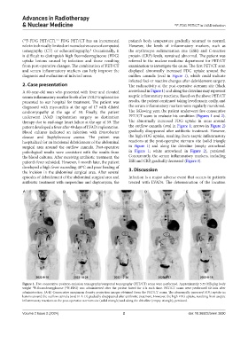
Figure 1. Five consecutive positron emission tomography/computed tomography (PET/CT) scans were performed. Approximately 5.55 MBq/kg body
weight F-fluorodeoxyglucose ( F-FDG) was administered after the patient fasted for 4 h each time. PET/CT scans were performed 60 min after
18
18
administration. (A-E) Consecutive maximum density projection images obtained from the PET/CT scans. The abnormally increased FDG uptake in
lesions around the outflow cannula (oval in A-D) gradually disappeared after antibiotic treatment. However, the high FDG uptake, resulting from aseptic
inflammatory reactions at the post-operative sternum site (solid triangle) and along the driveline (empty triangle), persisted.
Volume 2 Issue 2 (2024) 2 doi: 10.36922/arnm.3690