
Page 25 - ARNM-2-4
P. 25
Advances in Radiotherapy
& Nuclear Medicine 68 Ga-PSMA PET CT/MRI in prostate cancer
for invasive biopsies. In Figure 2, it demonstrates high the image exhibits high PSMA uptake, demonstrating its
diagnostic accuracy in identifying primary lesions, lymph effectiveness in detecting primary lesions, lymph node
node involvement, and distant metastases, aiding in TNM involvement, and distant metastases.
staging and treatment planning. 68 Ga-PSMA-PET/CT not only aids in staging but also helps
68 Ga-PSMA-PET, particularly when combined with predict nodal involvement and tumor aggressiveness, thereby
CT or MRI, has shown high diagnostic accuracy in TNM informing treatment strategies and potentially reducing
staging of PCa, with notable sensitivity in detecting unnecessary surgical interventions. There is a significant
primary lesions and superior performance in identifying association between PSMA uptake and GS, indicating that
lymph nodes and distant metastases. As shown in Figure 3, 68 Ga-PSMA PET can serve as a predictive tool for tumor
aggressiveness and disease prognosis. Higher SUV values
max
correlate with elevated GS and PSA levels, suggesting that
68 Ga-PSMA-PET findings can reflect the severity of PCa and
aid in stratifying patients based on their risk profiles.
5. Conclusion
The incorporation of Ga-PSMA-PET, particularly when
68
integrated with MRI, holds significant promise for the initial
diagnosis and management of PCa. Its ability to correlate
with key clinical parameters and improve diagnostic
accuracy underscores its potential as a valuable tool in
clinical practice. However, addressing current limitations
through standardization and further research is essential to
fully realize the benefits of PSMA-PET in PCa care.
Acknowledgments
Figure 2. Comparison of Ga-PSMA PET/CT (left) and Ga-PSMA-
68
68
PET (right) scans for staging prostate cancer. The images demonstrate None.
high diagnostic accuracy in identifying primary lesions, lymph node
involvement, and distant metastases, aiding in TNM staging and Funding
treatment planning.
Abbreviations: Ga-PSMA-PET: Gallium-68-prostate-specific membrane None.
68
antigen-positron emission tomography; CT: Computed tomography.
Conflict of interest
Kalevi Kairemo is the Editorial Board Member of this journal,
but was not in any way involved in the editorial and peer-
review process conducted for this paper, directly or indirectly.
Separately, other authors declared that they have no known
competing financial interests or personal relationships that
could have influenced the work reported in this paper.
Author contributions
Conceptualization: Philip F. Cohen, Kalevi Kairemo
Writing – original draft: I.O. Akinmuleya
Writing – review & editing: All authors
Ethics approval and consent to participate
Not applicable.
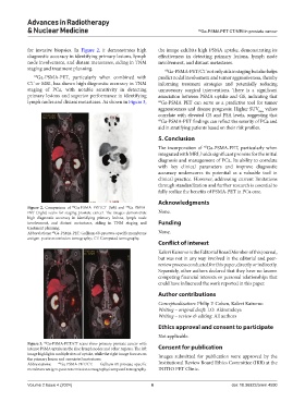
Figure 3. Ga-PSMA-PET/CT scans show primary prostate cancer with
68
intense PSMA uptake in the iliac lymph nodes and other regions. The left Consent for publication
image highlights multiple sites of uptake, while the right image focuses on Images submitted for publication were approved by the
the primary lesion and associated metastases.
Abbreviations: 68 Ga-PSMA-PET/CT: Gallium-68-prostate-specific Institutional Review Board Ethics Committee (IRB) at the
membrane antigen-positron emission tomography/computed tomography. INITIO PET Clinic.
Volume 2 Issue 4 (2024) 6 doi: 10.36922/arnm.4590