
Page 34 - ARNM-2-4
P. 34
Advances in Radiotherapy
& Nuclear Medicine 3D-PT-assisted CT-guided I RSI
125
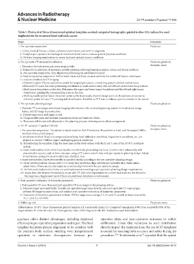
Table 1. Protocol of three‑dimensional‑printed template‑assisted computed tomography‑guided iodine‑125 radioactive seed
implantation for recurrent head and neck cancer
Steps Executors
1. Pre-operative assessment Physician
i. Collect medical history, conduct a physical examination, and confirm diagnosis.
ii. Complete pre-operative hematological and biochemical tests to evaluate general physical conditions.
iii. Perform imaging examinations to assess local and systemic tumor conditions.
2. Pre-operative CT-simulated localization Physician, physicist,
i. Discussion for indications and assess surgical risks therapist, nurse
ii. Prepare for localization. If necessary, provide patient positioning training in supine, prone, and lateral positions.
iii. Pre-operative preparation: Skin disinfection following the established protocol.
iv. Positioning device preparation: Utilize mesh masks and body vacuum cushions for combined fixation techniques.
v. Location simulated by CT imaging:
• Position fixation: Choose a position suitable for surgical procedures, considering patient comfort and tolerance.
• Enhanced CT scans: Use laser positioning coordinates to mark surface entry, exit, and left and correct laser line positions.
• Mark tumor boundaries on the skin: Determine the upper and lower tumor boundaries and the left and right tumor
boundaries, marking the corresponding areas on the skin.
• Marking needle points: Select the tumor center as the fixed needle channel design point or choose bony structures as
reference points for easy CT scanning and identification. Establish an X-Y axis coordinate system centered on the tumor.
3. Pre-operative planning design Physician, physicist
i. Transmit CT scan images and related imaging information to the treatment planning system for localization, image
fusion, and 3D image reconstruction.
ii. Outline target areas and organs at risk.
iii. Design needle paths and establish prescription doses and limits for OARs.
iv. Physicians collaborate with physicists in the planning process, obtaining senior physician approval.
4. 3D-PT-assisted CT-guided 125I RSI Physician, physicist,
i. Pre-operative preparation: The patient is repositioned on the CT simulator, the position is fixed, and the surgical field is therapist, nurse
locally disinfected and draped.
ii. Anesthesia method: Deep vein compound anesthesia, local infiltration anesthesia, lingual nerve anesthesia, etc., are
adopted as needed. Children require undergoing general anesthesia.
iii. Repositioning the template: Align the laser lines on the body surface with the X and Y axes of the 3D-PT coordinate
system.
iv. Insert fixed needles: Insert three fixed needles into the body, penetrating 2 to 3 cm. Confirm their relationship with
anatomical structures, such as bone structure, using a CT scan to match them with pre-operative planning design. Adjust
for deviations, aiming for an error of≤2 mm, ideally≤1 mm.
v. Insert seed needles: Insert seed needles to specified depths according to the pre-operative planning design.
vi. Verify needle positions: Rescan with CT to verify that needle tips align with the pre-operative plan. Make minor
adjustments if there are any discrepancies to ensure alignment with the pre-operative design.
vii. Perform seed implantation: Carry out seed implantation according to pre-operative planning design requirements.
viii. Assess dose distribution: Immediately rescan with CT after seed implantation to confirm that seeds are distributed in
the target area. Supplement seeds if there are positional deviations or movements.
5. Post-operative evaluation of dosimetry parameters Physician, physicist
i. Post-operative CT scan: Transmit post-operative CT scan images to the planning system.
ii. Delineate target areas and OARs: Transfer pre-operative target areas directly onto post-operative CT scan images,
perform 3D image reconstruction, and conduct post-operative evaluation of dosimetric parameters.
iii. Evaluation of dosimetric parameters includes D90 for target area coverage, CI, HI, and EI, as well as doses received by
0.1, 1, and 2 cc of OARs.
6. Follow-up visit Physician, nurse
Abbreviations: 3D-PT: Three-dimensional-printed template; CI: Conformity index; CT: Computed tomography; D90: Dose received by 90% of the
target volume: EI: External index; HI: Homogeneity index; OAR: Organs at risk; RSI: Radioactive seed implantation.
guidance offers distinct advantages, including improved operative plans and post-operative outcomes to within
efficiency in pre-operative preparation and repair. The fixed millimeters. Given that variations in seed distribution
template facilitates precise alignment of its crosshair with directly impact the radiation dose, the use of 3D templates
the patient’s body surface, enabling even inexperienced is crucial for ensuring both accuracy and safety during the
operators to minimize discrepancies between pre- procedure, 35,36 Rembowska et al. reported that the spatial
37
Volume 2 Issue 4 (2024) 7 doi: 10.36922/arnm.4212