
Page 101 - ARNM-3-1
P. 101
Advances in Radiotherapy
& Nuclear Medicine PET/CT in B-cell non-Hodgkin’s lymphoma
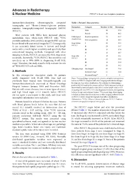
immunohistochemistry, ultrasonography, computed Table 1. Patients’ characteristics
tomography, and 18 fluoro-2-deoxy-d-glucose positron
emission tomography/computed tomography (18FDG Parameters Category Number (n=86) Percentage
PET/CT). Sex Male 48 55.8
Female 38 44.2
Most patients with NHLs have increased glucose
metabolism; thus, whole-body PET/CT with 18 FDG Pathological Rapid progression 72 83.7
classification
provides molecular-level metabolic images by PET and can Slow progression 14 16.3
be combined with anatomical images by CT. Consequently, Stage before I 26 30.2
it can accurately detect lesions in tumors and lymph PET/CT II 29 33.7
nodes with a much higher sensitivity and specificity than III 15 17.4
conventional imaging methods. Compared with other IV 16 18.7
imaging methods, PET/CT can assess the NHL stage more Stage after I 19 22.1
accurately. Reportedly, FDG PET/CT has sensitivity and PET/CT
18
specificity up to 99%–100% in diagnosing B-cell NHL II 23 26.8
stage. Therefore, this study aimed to fully evaluate the role III 18 20.9
2
of FDG PET/CT in B-cell NHL. IV 26 30.2
18
Changing stage Unchanged 65 75.6
2. Methods Increased stage* 21 24.4
In this retrospective descriptive study, 86 patients Decreased stage** 0 0
newly diagnosed with B-cell NHL who had not Notes: *Increased stage: upstaging is the presence of additional suspicious
previously been treated were histopathologically and lesions on PET/CT compared with other imaging methods that changes
immunohistochemically examined at Hanoi Oncology the stage per Ann Arbor classification. The criterion for determining a
18
Hospital between January 2018 and December 2022. positive PET/CT result is the presence of focal FDG increased uptake as
determined by standard uptake value (SUV) and/or lymph nodes on CT
Patients with severe diseases, two or more types of cancer, measuring ≥10 mm. SUV of 2.5 is the diagnostic threshold, distinguishing
and high blood sugar ≥11.1 mmol/L before PET/CT, between benign and malignant lesions. **Decreased stage: downstaging
did not agree to participate in the study, and those with is defined as lesions that are suspicious on other imaging methods but not
incomplete information were excluded. on PET/CT, which change the stage per Ann Arbor classification.
18
Abbreviation: FDG: 18fluoro-2-deoxy-d-glucose; PET/CT: Positron
Patients fasted for at least 6 h before the scan. Patients emission tomography/computed tomography.
with blood glucose levels before the scan that did not
exceed 11.1 mmol/L received an intravenous dose of The PET/CT stages before and after the procedure
18 FDG of 0.14 – 0.15 mCi/kg. After FDG injection, differed (Table 1). The number of patients in Stages III and
18
patients rested in the waiting room. After 45 – 60 min, IV increased after PET/CT compared with that before the
patients underwent full-body PET/CT using the GE scan. The Stage III rate increased to 20.9%, particularly Stage
PET/CT system. The results were processed using IV, which remarkably increased to 30.2%. After PET/CT,
specialized software, read, and agreed on by two nuclear the stage increased in 24.4% of the patients and remained
medicine specialists. Information about age, sex, pathology the same in 75.6%; none of the patients had a lowered stage.
type, lesion location, lesion size injury, and maximum After PET/CT, the patients were distributed as follows:
standard uptake value of the injury were recorded. three patients from stage I were reassigned to Stage II,
The data were analyzed using IBM SPSS Statistics three from Stage I to Stage III, one from Stage I to Stage IV,
version 20 (IBM Corp., Armonk, NY, USA). Normally five from Stage II to Stage III, four from Stage II to Stage
distributed quantitative variables were expressed as IV, and five from Stage III to stage IV. None of the patients
mean ± standard deviation and non-normally distributed lowered the stage (Figure 1).
variables as median. The t- and Mann–Whitney tests were The post-PET/CT increase rate of the slow-progressing
used to compare two means and medians, respectively. histopathological group was lower (21.4%) than that of the
3. Results rapid-progressing histopathological group (25%). However,
this difference was not significant with P > 0.05 (Table 2).
Patient characteristics are summarized in Table 1.
4. Discussion
A total of 86 patients were recruited, of whom 55.8%
were men. The median age of the patients was 58.1 years. For B-cell NHL, accurate determination of disease stage
Most patients had rapidly progressive pathology (n = 72). is crucial for selecting appropriate treatment methods,
Volume 3 Issue 1 (2025) 93 doi: 10.36922/arnm.4813