
Page 102 - ARNM-3-1
P. 102
Advances in Radiotherapy
& Nuclear Medicine PET/CT in B-cell non-Hodgkin’s lymphoma
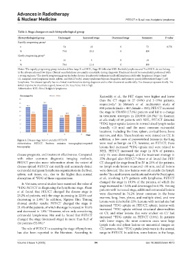
Table 2. Stage changes on each histopathological group
Histopathological group Unchanged Increased stage Decreased stage Summary P‑value
Rapidly progressing group a
n 54 18 0 72 0.776
% 75.0 25.0 0 100
Slowly progressing group b
n 11 3 0 14
% 78.6 21.4 0 100
Notes: The rapidly progressing group includes diffuse large B-cell NHL, Stage III follicular NHL. Burkitt’s lymphoma and T-cell NHL do not belong
a
to the aforementioned two types. Clinical manifestations are usually remarkable during diagnosis. Treatment should be immediately performed with
a strong regimen. The slowly progressing group includes chronic lymphocytic leukemia/small cell lymphoma, follicular lymphoma (stages I and
b
II), marginal zone lymphoma (node, spleen, and MALT body), sezary syndrome/Mycosis fungoides, and primary poorly differentiated large T-cell
lymphoma. The disease typically has no clinical manifestations during diagnosis and is often discovered accidentally. The disease progresses slowly. The
initial response to treatment is good; however, the recurrence risk is high.
Abbreviation: NHL: Non-Hodgkin’s lymphoma.
Kasireddy et al., the PET stages were higher and lower
than the CT stages in 27 (14%) and 3 (1%) patients,
respectively. In Metser’s et al. multicenter study of
6
850 patients (male = 467; female = 383), PET/CT increased
the stage in 150/850 (17.6%) patients and led to a change
in treatment strategies in 224/850 (26.4%). In Raanani
7
et al.’s study of 68 patients with NHL, PET/CT detected
18 FDG hyper-uptake lesions in normal-sized lymph nodes
(usually <10 mm) and the most common extranodal
locations, including the liver, spleen, cortical bone, bone
marrow, and skin. These locations were missed on CT. In
Figure 1. Disease stage before and after PET/CT addition, a few cases of paravertebral lesions in the lung
Abbreviation: PET/CT: Positron emission tomography/computed were read as benign on CT; however, on PET/CT, these
tomography. lesions had increased FDG uptake and were related to
18
NHL. PET/CT increased the stage in 31% of patients,
disease prognosis, and treatment effectiveness. Compared only 1% were downstaged, and the treatment strategy in
with other common diagnostic imaging methods, 25% changed after PET/CT. Omar et al. found that PET/
8
PET/CT provides more information about the extent of CT changed the stage from II to IV in 25% of the patients,
disease spread. PET/CT can rapidly and accurately detect no lymph node lesions measured <10 mm, and all lesions
extranodal malignant lymphoma organizations in the liver, were detected. The new lesions were all outside the lymph
spleen, soft tissue, etc., due to the higher-than-normal nodes. In a multicenter, multinational study by Barrington
9
absorption of FDG of these organizations. et al., involving 1,171 patients with lymphoma, PET/CT
18
changed the stage in 19.9% of the patients, of which the
In Vietnam, several studies have examined the value of
18 FDG PET/CT in diagnosing the lymphoma stage. Pham stage increased in 13.6% and downstaged in 6.3%. Among
et al. found that PET/CT changed the disease stage in patients with increased stage, additional extranodal lesions
were discovered in 74.2% (most common in the bone
25.2% of patients, with the stage increasing in 22.8% and
decreasing in 2.4%. In addition, Nguyen Huu Thuong marrow, lung, liver, pleura, and multiple sites), and new
3
showed similar results: PET/CT changed the stage in lesions were detected in 22%. Lesions with normal size but
increased FDG uptake on PET/CT, splenic lesions with
18
21.4% of the patients, of which the stage increased in 19.6% increased FDG uptake without structural abnormalities
18
and decreased in 1.8%. However, when only considering on CT, and other lesions that were unclear on CT had
4
extranodal lymphomas, Mai and Le found that PET/CT increased FDG uptake on PET/CT (3.8%). In patients
18
changed the stage (increased stage) in more than half of with lower stages, the most common cases were large
the patients (52.6%). 5
lymph node and spleen lesions on anatomical images on
The role of PET/CT in assessing the stage of lymphoma CT; however, their FDG uptake levels were in the normal
18
has also been reported in the literature. According to range in PET/CT. In addition, some lesions in the lungs,
Volume 3 Issue 1 (2025) 94 doi: 10.36922/arnm.4813