
Page 107 - ARNM-3-1
P. 107
Advances in Radiotherapy
& Nuclear Medicine Role of 18F-FDG in brown tumor detection
A B
C
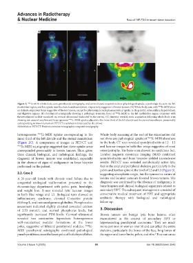
Figure 2. 99m Tc-MIBI whole-body scan, parathyroid scintigraphy, and cervical static acquisition show physiological uptake, a pathologic focus in the left
mediolobar region, and low uptake near the clavicle and manubrium, respectively, suggestive of brown tumors. (A) Whole-body scan with Tc-MIBI show
99m
no definite suspicious focus suggestive of brown tumors, except for physiological radiopharmaceutical uptake in the parotid, submaxillary, hepatobiliary,
and digestive regions. (B) Parathyroid scintigraphy showing a pathologic retention focus of 99m Tc-MIBI in the left mediolobar region consistent with
the retrothyroid nodule visualized on cervical ultrasound (indicated by the arrow). (C) Anterior cervical static acquisition following whole-body scan
showing two areas of very low and heterogeneous Tc-MIBI uptake adjacent to the inner third of the left clavicle and the sternal manubrium, presumably
99m
corresponding to brown tumors on PET/CT examination (indicated by the arrow).
Abbreviation: PET/CT: Positron emission tomography/computed tomography.
heterogeneous 99m Tc-MIBI uptake corresponding to the Whole-body scanning at the end of the examination did
99m
inner third of the left clavicle and the sternal manubrium not show any pathological uptake of Tc-MIBI elsewhere
(Figure 2C). A comparison of images in PET/CT and in the body. CT scan revealed spondylodiscitis at L2 – L3
99m Tc-MIBI scintigraphy suggested that these uptake areas and lacunar images in both iliac wings suggestive of renal
corresponded presumably to brown tumors. Thus, given osteodystrophy. The bone scan showed no suspicious foci.
these clinical, biological, and radiological findings, the Lumbar magnetic resonance imaging (MRI) confirmed
diagnosis of brown tumors was established, especially spondylodiscitis and bone biopsies yielded inconclusive
in the absence of signs of malignancy on bone biopsies results. PET/CT scan revealed metabolically active lytic
performed on the patient. foci in the axial and peripheral skeleton, particularly in the
pelvis and lumbar spine at the level of L2 and L3 (Figure 3),
2.2. Case 2 suggesting a neoplastic origin, but the symmetric nature of
A 21-year-old female with chronic renal failure due to lesions and lacunar contours favored brown tumors. The
congenital urological malformation presented to the diagnosis was confirmed by the absence of malignancy in
rheumatology department with pelvic pain, lumbalgia, bone biopsies and clinical-biological arguments related to
and weight loss. X-rays revealed lytic lacunar images secondary HPT. The subsequent management consisted of
in both iliac wings and L3. Biological tests showed an conservative medical treatment of HPT and appropriate
inflammatory syndrome, elevated C-reactive protein antibiotic therapy with biological and radiological
60.8 mg/L, and increased gamma globulins. Phosphocalcic follow-up.
assessment indicated slightly elevated corrected calcium
of 2.53 mmol/L and normal phosphorus levels, but 3. Discussion
significantly increased PTH levels. Cervical ultrasound Brown tumors are benign lytic bone lesions, often
revealed two centimetric hypoechoic heterogeneous encountered in the context of secondary HPT or
well-vascularized nodular formations at the lower hypersecreting parathyroid carcinoma. They are much
1
poles, suggestive of bilateral parathyroid nodules. 99m Tc- more common in women over 50 and can affect the entire
MIBI parathyroid scintigraphy confirmed pathological skeleton, particularly the bones of the face, long bones of
parathyroid tissue near the lower poles of both thyroid lobes. the upper and lower limbs, pelvis, and ribs. They can cause
2
Volume 3 Issue 1 (2025) 99 doi: 10.36922/arnm.3540