


Page 103 - ARNM-3-3
P. 103
Advances in Radiotherapy
            & Nuclear Medicine                                                             Sarcomatoid HCC therapy


              Several clinical trials have suggested that transarterial   Mai Hospital diagnosed the tumor as SHC with cirrhosis
            chemoembolization (TACE), radiofrequency ablation   and 80% tumor necrosis (Figure 2). The patient recovered
            (RFA), and percutaneous ethanol injection (PEI) may   postoperatively and was discharged for follow-up.
            contribute to the transformation from HCC to SHC.    In September 2023, during a routine follow-up,
                                                          1
            However, SHC has also been reported in patients    an abdominal CT scan revealed multiple masses and
            without a history of these treatments, indicating that its   nodules of heterogeneous density with variable contrast
            exact pathogenesis remains unclear. At present, there   enhancement in the greater omentum close to the left
            is no established effective treatment for SHC. Surgical   anterior abdominal wall, the largest measuring 47 × 34 mm
            resection remains the primary therapeutic approach, with   (Figure 3A). A well-defined nodule measuring 25 × 18 mm
            additional regional and systemic therapies; however, their   with  poor  enhancement  was observed in  segment
            efficacy in improving survival outcomes has not been well   VIII of the liver parenchyma (Figure  3B). This nodule
            demonstrated.                                      exhibited the typical trabecular morphology of HCC on
              We  herein  present  a  clinical  case  of  a  male  patient   histopathological examination (Figure 4).
            diagnosed with SHC who experienced multiple recurrences   The patient was diagnosed with recurrent SHC and
            and underwent multimodal treatment, including TACE,   subsequently underwent colectomy, small intestine
            surgery, and systemic chemotherapy.                resection,  and  PEI  (Figure  5).  Immunohistochemical

            2. Case presentation
                                                                A                     B
            A 57-year-old male patient with a history of hepatitis B,
            treated for 10 years but now off treatment, was diagnosed
            with HCC in 2013 and underwent TACE five times at Bach
            Mai Hospital, Hanoi, Vietnam.
              In April 2023, the patient was diagnosed with recurrent
            HCC and underwent a sixth TACE at Duc Giang General
            Hospital, Hanoi, Vietnam. He was discharged for follow-up
            after treatment.

              In May 2023, the patient was admitted to the hospital
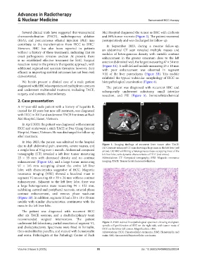
            due to dull abdominal pain, anorexia, severe nausea, and   Figure  1. Imaging findings of recurrent liver tumor after TACE.
                                                               (A) Contrast-enhanced CT scan showing a large mass in the left liver (red
            a weight loss of 5 kg over 1 month. Abdominal computed   arrow). (B) MRI exhibiting a heterogeneous mass occupying most of the
            tomography (CT) revealed a left liver tumor measuring   left liver lobe, with dynamic characteristics of HCC (red arrow).
            25 ×  35  mm with decreased density  and no contrast   Abbreviations: CT: Computed tomography; MRI: Magnetic resonance
            enhancement (Figure 1A), and a large tumor measuring   imaging; TACE: Transarterial chemoembolization.
            97 × 145  mm occupying almost the entire left liver
            lobe, with characteristics suggestive of HCC. Magnetic
            resonance imaging (MRI) showed a localized mass in
            segment VI measuring 40 × 39 × 24 mm with no contrast
            enhancement. Adjacent to the left liver lobe, there was
            a large heterogeneous mass measuring 94 × 153  mm,
            exhibiting central and peripheral necrosis, arterial-phase
            contrast enhancement, and venous phase wash-out
            (Figure 1B). In addition, segment II had a 28 × 18 × 20 mm
            nodule with similar characteristics, continuous with the
            mass in the left liver lobe.
              The patient was diagnosed with recurrent HCC
            after  six  TACE  sessions,  and  a  multidisciplinary  team
            recommended surgical intervention. The patient
            underwent left lobectomy, partial resection of segment VI,   Figure 2. H&E-stained histopathological specimen showing malignant
            and cholecystectomy. Specimens were  fixed  in formalin,   spindle cell proliferation of SHC on the right side, with tumor nests of
                                                               HCC on the lower left corner. Magnification: ×100.
            then embedded in paraffin, and stained with hematoxylin   Abbreviations: HCC: Hepatocellular carcinoma; H&E: Hematoxylin and
            and eosin. Pathologists at the Pathology Center of Bach   eosin; SHC: Sarcomatoid hepatocellular carcinoma.


            Volume 3 Issue 3 (2025)                         95                        doi: 10.36922/ARNM025220024

