


Page 104 - ARNM-3-3
P. 104
Advances in Radiotherapy
            & Nuclear Medicine                                                             Sarcomatoid HCC therapy


             A                      B


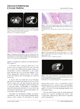
                                                               Figure 5. H&E-stained histopathological features of metastatic HCC to
                                                               the colon, showing tumor cells resembling hepatocytes within the colonic
                                                               wall. Magnification: ×40.
            Figure 3. CT imaging findings of recurrent tumor. (A) Multiple masses   Abbreviations: HCC: Hepatocellular carcinoma; H&E: Hematoxylin and
            and nodules in the greater omentum (red arrow). (B) A hypodense   eosin.
            nodule in the right liver parenchyma (red arrow).
            Abbreviation: CT: Computed tomography.


                                                               Figure  6.  Immunohistochemical  staining  with HepPar-1  is  a  useful
            Figure 4. H&E-stained histopathological specimen showing trabecular   tool to confirm the hepatic origin of metastatic tumors in the colon.
            morphology of HCC after TACE at low magnification. Magnification: ×40  Magnification: ×100.
            Abbreviations: HCC: Hepatocellular carcinoma; H&E: Hematoxylin and   Abbreviation: HerPar-1: Hepatocyte paraffin 1.
            eosin; TACE: Transarterial chemoembolization.

            analysis of postoperative specimens confirmed metastatic
            HCC (Figure 6).
              In  November  2023,  a  follow-up  abdominal  CT  scan
            showed a mass  in segment VIII of the  liver measuring
            84 × 70 mm, with a well-defined boundary, strong, and
            heterogeneous contrast enhancement in the arterial
            phase, and wash-out in the venous phase (Figure 7). The
            patient subsequently underwent another TACE session
            at Bach Mai Hospital and was discharged for continued   Figure  7.  Axial  contrast-enhanced  CT  image  showing  a  right  hepatic
            monitoring.                                        mass with characteristics of HCC (red arrow), demonstrating an increase
                                                               in size compared to the September 2023 scan
              In  January  2024,  the  patient  had  severe  abdominal   Abbreviations: CT: Computed tomography; HCC: Hepatocellular carcinoma.
            pain and was admitted for an abdominal CT scan, which
            showed a large mass with clear boundaries measuring   intravenously on days 1 and 2; (ii) ifosfamide 3.75 g/m
                                                                                                             2
            130 × 94 × 108 mm in the right liver lobe, including areas   intravenously over 4 h on days 1 and 2; and (iii) Mesna
            of increased density corresponding to interventional   750 g/m  intravenously before each ifosfamide cycle, then
                                                                      2
            material and decreased density due to necrosis, with strong   again at 4 h and 8 h after the ifosfamide infusion. Each
            heterogeneous arterial-phase contrast enhancement, and   cycle lasted 21 days.
            venous phase wash-out (Figure 8).
                                                                 After two chemotherapy cycles, the patient developed
              The patient was subsequently initiated on systemic   elevated liver enzymes, prompting the medical team
            chemotherapy  with  the  anthracycline-ifosfamide-mesna   to discontinue chemotherapy and switch to supportive
            regimen, which included (i)  doxorubicin 30  mg/m    medical management.
                                                          2

            Volume 3 Issue 3 (2025)                         96                        doi: 10.36922/ARNM025220024

