


Page 16 - ARNM-3-3
P. 16
Advances in Radiotherapy
            & Nuclear Medicine                                              Review of image-guided adaptive radiotherapy


                                                               Table 2. Main parameters of the uRT‑506c platform
                                                               Feature                      Content
                                                               Layout           Integrated design, diagnostic 16-row spiral
                                                                                CT, and linear accelerator co-bed coaxial
                                                                                design
                                                               CT aperture      70 cm diameter
                                                               The angle of rotation of   −362° – 182° and maximum rotation speed
                                                               the accelerator frame  of 7°/s
                                                               Beams            6 MV (FF/FFF) X-ray
                                                               Dose rate        25 – 600 MU/min (FF), 75 – 1400 MU/min
                                                                                (FFF)
                                                               Multi-leave collimators  1.0 cm×20 pair, 0.5 cm×40 pair, velocity of
                                                                                the lobes 2.5 cm/s
                                                               The radiation field  40×40 cm 2
                                                               Radiotherapy equipment 3D-CRT, IMRT, uARC
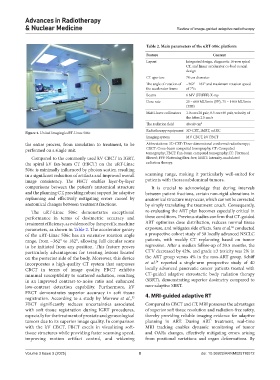
            Figure 4. United Imaging’s uRT-Linac 506c
                                                               Imaging system   MV CBCT, kV FBCT
            the entire process, from simulation to treatment, to be   Abbreviations: 3D-CRT: Three-dimensional conformal radiotherapy;
            performed on a single unit.                        CBCT: Cone-beam computed tomography; CT: Computed
                                                               tomography; FBCT: Fan-beam computed tomography; FF: Flattened
              Compared to the commonly used kV CBCT in IGRT,   filtered; FFF: Flattening filter-free; IMRT: Intensity-modulated
            the spiral kV fan-beam CT (FBCT) on the uRT-Linac   radiation therapy.
            506c is minimally influenced by photon scatter, resulting
            in a significant reduction of artifacts and improved overall   scanning range, making it particularly well-suited for
            image consistency. The FBCT enables layer-by-layer   patients with thoracoabdominal tumors.
            comparisons  between  the  patient’s  anatomical  structure   It is crucial to acknowledge that during intervals
            and the planning CT, providing robust support for adaptive   between patient fractions, certain non-rigid alterations in
            replanning  and effectively  mitigating errors caused  by   anatomical structure may occur, which cannot be corrected
            anatomical changes between treatment fractions.    by simply translating the treatment couch. Consequently,

              The uRT-Linac 506c demonstrates exceptional      re-evaluating the ART plan becomes especially critical in
            performance in terms of dosimetric accuracy and    these conditions. Previous studies confirm that CT-guided
            treatment efficiency, as evidenced by the specific machine   ART optimizes dose distribution, reduces normal tissue
                                                                                                   53
            parameters, as shown in Table 2. The accelerator gantry   exposure, and mitigates side effects. Sara et al.  conducted
            of the uRT-Linac 506c has an extensive rotation angle   a prospective cohort study of 50 locally advanced NSCLC
            range, from −362° to 182°, allowing full circular scans   patients,  with  weekly  CT  replanning  based  on  tumor
            to  be  initiated  from  any position.  This  feature proves   regression. After a median follow-up of 20.5 months, the
            particularly advantageous for treating lesions located   CTV decreased by 42%, and grade ≥3 toxicity was 2% in
            on the posterior side of the body. Moreover, this device   the ART group versus 4% in the non-ART group. Schiff
                                                                   54
            incorporates a high-quality CT system that surpasses   et al.  reported a single-arm prospective study of 40
            CBCT in terms of image quality. FBCT exhibits      locally advanced pancreatic cancer patients treated with
            minimal susceptibility to scattered radiation, resulting   CT-guided adaptive stereotactic body radiation therapy
            in  an  improved  contrast-to-noise ratio and enhanced   (SBRT), demonstrating superior dosimetry compared to
            low-contrast detection capability. Furthermore, kV   non-adaptive SBRT.
            FBCT demonstrates superior accuracy in soft tissue   4. MRI-guided adaptive RT
            registration. According to a study by Morrow  et  al.,
                                                         52
            FBCT significantly reduces uncertainties associated   Compared to CBCT and CT, MRI possesses the advantages
            with  soft  tissue registration during IGRT procedures,   of superior soft tissue resolution and radiation-free safety,
            especially for the treatment of prostate and gynecological   thereby providing reliable imaging evidence for adaptive
            tumors due to its superior image quality. In comparison   planning  in ART. During ART treatment, real-time
            with the kV CBCT, FBCT excels in visualizing soft-  MRI tracking enables dynamic monitoring of tumor
            tissue structures while providing faster scanning speed,   and OARs changes, effectively mitigating errors arising
            improving motion artifact control, and widening    from positional variations and organ deformations. By


            Volume 3 Issue 3 (2025)                         8                          doi: 10.36922/ARNM025110012

