


Page 12 - ARNM-3-3
P. 12
Advances in Radiotherapy
            & Nuclear Medicine                                              Review of image-guided adaptive radiotherapy


            on computed tomography (CT) and other imaging      a specific margin to generate a new planning target volume
            modalities such as positron emission tomography-CT and   (PTV), ensuring adequate radiation dose delivery to the
            magnetic resonance imaging (MRI), to optimize treatment   tumor. However, this approach can only partially enhance
            precision and effectiveness. 6-11  During RT, technicians use   tumor coverage while escalating radiation exposure to
            auxiliary equipment, such as positioning laser lights, to   surrounding normal tissues, thereby constraining the
            ensure precise alignment between the irradiation site and   feasibility of administering higher therapeutic doses to
            the planned target. This approach minimizes the risk of   the target lesion. Moreover, the determination of CTV
            misdirected radiation or excessive doses to organs at risk   expansion  distance  is  typically  based  on  fixed  standards
            (OARs) resulting from target movement and changes in   derived from clinical research summaries, disregarding
            patient positioning.                               patient-specific anatomical variations. Consequently,
                                                               in certain cases, the expansion distance may be either
              However, considering the prolonged duration of RT,   excessive or  insufficient.  An excessively large expansion
            typically spanning approximately 30 – 40 days to complete   compromises protection of OARs and restricts the
            the entire course, several factors may change during this   potential for target dose escalation, whereas an inadequate
            period. These include alterations in the patient’s body   expansion may result in reduced target dose coverage. 16-21
            shape, tumor size and morphology, bladder or organ
            filling status, displacement of OARs adjacent to the target   To achieve an optimal equilibrium between target
                                                                                                         22
            area, and variations in beam entry distances. 12-15  Such   expansion distance and radiation dosage, Yan et al.  and
                                                                       23
            alterations can cause discrepancies between the patient’s   Yan  et al.  proposed the innovative concept of adaptive
            anatomy at the time of each treatment fraction and the   RT (ART). ART is a closed-loop radiation treatment
            anatomical model used for treatment planning, potentially   process where the treatment plan can be modified using
            compromising the accuracy of dose delivery. These effects   systematic feedback of measurements. ART intends to
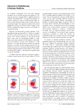
            are illustrated in Figure 1, which presents examples from   improve radiation treatment by systematically monitoring
            various RT modalities.                             treatment variations and incorporating them to re-optimize
                                                               the treatment plan early on during the treatment course.
              To address this issue, clinicians commonly employ a   In this process, field margin and treatment dose can be
            strategy of expanding the clinical target volume (CTV) by   routinely customized to each patient to achieve a safe dose
                                                               escalation. By individually optimizing prescribed dosage
            A                                                  and expansion distance  for each patient, and adapting
                                                               initial treatment plans to account for anatomical variations
                                                               when necessary, ART aims to increase tumor local control
                                                               rates and decrease toxicity of the OARs. 24-29
                                                                 The ART approach primarily comprises three modes:
                                                               (i) Offline ART utilizes offline imaging to adjust treatment
                                                               plans between fractions without requiring real-time
                                                               capabilities. Although being user-friendly, this mode has
                                                               limited capacity to evaluate actual dose delivery due to the
            B                                                  lack of same-day imaging information; 30-32  (ii) Online ART
                                                               employs online imaging to promptly complete target and
                                                               OAR delineation, re-optimize treatment plans, and deliver
                                                               treatment before each fraction. As the most widely used
                                                               mode in ART, it facilitates more effective assessment of
                                                               inter-fractional changes; however, further improvements
                                                               in optimization algorithms and integration processes are
                                                               still needed. 29,31,33-36 . The workflow of online ART is shown
                                                               in Figure 2. Artificial intelligence (AI), as the driving force
                                                               behind ART, has significantly enhanced the accuracy of
            Figure 1. Schematic diagram of anatomical structure variations between   automatic delineation for tumor targets and OARs through
            localization computed tomography and radiotherapy. (A) Anatomical   deep learning. It also enables intelligent processing of
            structure on the day of non-ART localization and corresponding   image reconstruction–such as synthetic CT generation,
            anatomy during treatment; (B) Anatomical structure on the day of ART
            radiotherapy localization and corresponding anatomy during treatment.  plan optimization, and quality assurance (QA)–thereby
            Abbreviations: ART: Adaptive radiotherapy; OAR: Organ at risk.  making real-time, dynamic adaptation feasible. Emerging


            Volume 3 Issue 3 (2025)                         4                          doi: 10.36922/ARNM025110012

