
Page 40 - BH-1-1
P. 40
Brain & Heart Epinephrine for CPVT
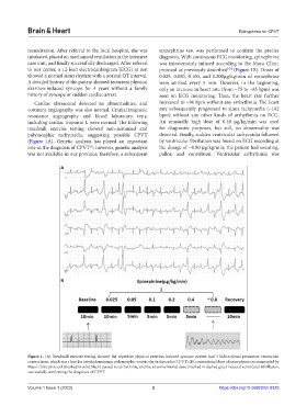
resuscitation. After referral to the local hospital, she was epinephrine test was performed to confirm the precise
intubated, placed on mechanical ventilation in the intensive diagnosis. With continuous ECG monitoring, epinephrine
care unit, and finally successfully discharged. After referral was intravenously infused according to the Mayo Clinic
to our center, a 12-lead electrocardiogram (ECG) at rest protocol as previously described [4,5] (Figure 1B). Doses of
showed a normal sinus rhythm with a normal QT interval. 0.025, 0.050, 0.100, and 0.200μg/kg/min of epinephrine
A detailed history of the patient showed recurrent physical were utilized every 5 min. However, in the beginning,
exertion-induced syncope for 4 years without a family only an increase in heart rate (from ~75 to ~85 bpm) was
history of syncope or sudden cardiac arrest. seen on ECG monitoring. Then, the heart rate further
Cardiac ultrasound detected no abnormalities, and increased to ~96 bpm without any arrhythmia. The heart
coronary angiography was also normal. Cranial magnetic rate subsequently progressed to sinus tachycardia (~102
resonance angiography and blood laboratory tests, bpm) without any other kinds of arrhythmia on ECG.
including cardiac troponin I, were normal. The following An unusually high dose of 0.40 μg/kg/min was used
treadmill exercise testing showed non-sustained and for diagnostic purposes, but still, no abnormality was
polymorphic tachycardia, suggesting possible CPVT detected. Finally, sudden ventricular tachycardia followed
(Figure 1A). Genetic analysis has played an important by ventricular fibrillation was found on ECG recording at
role in the diagnosis of CPVT ; however, genetic analysis the dosage of ~0.80 μg/kg/min; the patient had sweating,
[3]
was not available in our province; therefore, a subsequent pallor, and convulsion. Ventricular arrhythmia was
A
B
Figure 1. (A) Treadmill exercise testing showed the repetitive physical exertion-induced syncope patient had 3 bidirectional premature ventricular
contractions, which was a hint for catecholaminergic polymorphic ventricular tachycardia (CPVT).(B) conventional dose of epinephrine recommended by
Mayo Clinic protocol (marked in solid black) caused no arrhythmia, and the unconventional dose (marked in dashed grey) induced ventricular fibrillation,
successfully confirming the diagnosis of CPVT.
Volume 1 Issue 1 (2023) 2 https://doi.org/10.36922/bh.0325