
Page 63 - BH-2-2
P. 63
Brain & Heart Cerebral venous sinus assessment using MRI and CT
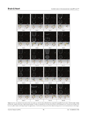
Figure A2. The case analysis in the present study. For each case, the left column illustrates the computed tomography-based results, while the right column
presents the magnetic resonance imaging-based analysis. The first row displays the coronary plane relative to the transverse sinus. The second row depicts the
transverse plane relative to the beginning of the sigmoid sinus. The third row showcases the three-dimensional geometric reconstructions based on the images.
Volume 2 Issue 2 (2024) 15 doi: 10.36922/bh.2756