
Page 62 - BH-2-2
P. 62
Brain & Heart Cerebral venous sinus assessment using MRI and CT
(Continued)
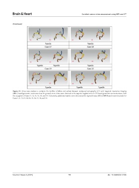
Figure A1. Sinus-wise analysis to compare the number of inlets and outlets between computed tomography (CT)-and magnetic resonance imaging
(MRI)-based geometric reconstructions. In general, more inlets were observed at the superior sagittal sinus in CT-based geometric reconstructions, with
the exception of Cases 7, 10, 15, 16, 18, and 19. Conversely, additional outlets were detected at the sigmoid sinus (SS) in MRI-based reconstructions for
Cases 1, 5, 7, 8, 9, 10, 13, 15, 16, 17, 18, and 19.
Volume 2 Issue 2 (2024) 14 doi: 10.36922/bh.2756