
Page 57 - BH-2-2
P. 57
Brain & Heart Cerebral venous sinus assessment using MRI and CT
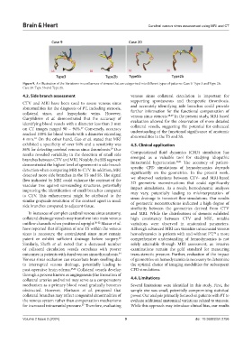
Figure 9. An illustration of the Variations in confluences of sinuses that are categorized into different types of patterns: Case 9. Type 3 and Type 2b;
Case 20. Type 5b and Type 2b.
4.2. Side branch assessment venous sinus collateral circulation is important for
CTV and MRI have been used to assess venous sinus supporting spontaneous and therapeutic thrombosis,
abnormalities for the diagnosis of PT, including stenosis, and accurately identifying side branches could provide
collateral sinus, and hypoplastic veins. However, further information for the functional compensation of
28,29
Guryildirim et al. demonstrated that the accuracy of venous sinus stenosis. In the present study, MRI-based
identifying blood vessels with a diameter less than 3 mm evaluation allowed for the observation of more detailed
collateral vessels, suggesting the potential for enhanced
on CT images ranged 90 – 94%. Conversely, accuracy understanding of the functional significance of anatomic
17
reached 100% for blood vessels with a diameter exceeding abnormalities in the TS and SS.
4 mm. On the other hand, Gao et al. stated that MRI
18
exhibited a specificity of over 94% and a sensitivity was 4.3. Clinical application
86% for detecting cerebral venous sinus thrombosis. Our
19
results revealed variability in the detection of small side Computational fluid dynamics (CFD) simulation has
branches between CTV and MRI. Notably, the SSS segment emerged as a valuable tool for studying idiopathic
30,31
demonstrated the highest level of agreement in side branch intracranial hypertension. The accuracy of patient-
detection when comparing MRI to CTV. In addition, MRI specific CFD simulations of hemodynamics depends
detected more side branches in the TS and SS. The signal significantly on the geometries. In the present work,
flow indicated by MRI could enhance the contrast of the we observed variations between CTV- and MRI-based
3D geometric reconstructions that could significantly
vascular tree against surrounding structures, potentially impact simulations. As a result, hemodynamic analyses
improving the identification of small branches compared may vary, potentially leading to misinterpretation of
to CTV. This enhancement might be attributed to the sinus drainage in transient flow simulations. Our results
similar grayscale resolution of the contrast agent in small of geometric reconstructions indicated a high degree of
side branches compared to adjacent tissue.
similarity between the geometries derived from CTV
In instances of complex cerebral venous sinus anatomy, and MRI. While the distributions of stenosis exhibited
collateral drainage vessels may transform into main venous high consistency between CTV and MRI, notable
outflow channels when positioned upright. 20-22 Mazur et al. variations were observed in anatomical parameters.
have reported that if ligation of one TS within the venous Although advanced MRI can visualize intracranial venous
sinus is necessary, the contralateral sinus must remain hemodynamics in patients with and without PT, a more
32
23
patent or exhibit sufficient drainage before surgery. comprehensive understanding of hemodynamics is not
Similarly, Sheth et al. noted that a decreased number solely attainable through MRI assessment, as invasive
of collateral circulation vessels correlates with poorer examinations remain the gold standard for measuring
outcomes in patients with dural venous sinus thrombosis. trans-stenotic pressure. Further, evaluation of the impact
24
Venous sinus occlusion can exacerbate brain swelling due of geometries on hemodynamics is necessary to determine
to interrupted venous drainage, potentially leading to the optimal choice of imaging modalities for subsequent
post-operative brain edema. 25,26 Collateral vessels develop CFD simulations.
through a process known as angiogenesis (the formation of
collateral arteries and veins) may serve as a compensatory 4.4. Limitations
mechanism as a primary blood vessel gradually becomes Several limitations were identified in this study. First, the
obstructed. However, Florisson et al. proposed that sample size was small, potentially compromising statistical
collateral branches may reflect congenital abnormalities of power. Our analysis primarily focused on patients with PT to
the venous system rather than compensation mechanisms evaluate additional anatomical variations related to stenosis.
for increased intracranial pressure. Therefore, evaluating While this approach may introduce clinical bias, our results
27
Volume 2 Issue 2 (2024) 9 doi: 10.36922/bh.2756