
Page 56 - BH-2-2
P. 56
Brain & Heart Cerebral venous sinus assessment using MRI and CT
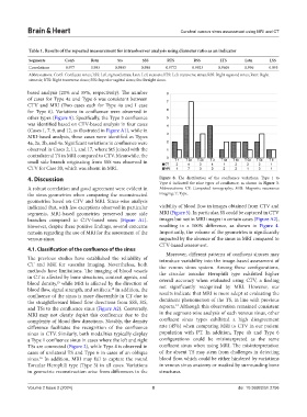
Table 1. Results of the repeated measurement for intraobserver analysis using diameter ratio as an indicator
Segments ConS Rstn Sts SSS RTS RSS LTS Lstn LSS
Correlations 0.977 0.993 0.9805 0.988 0.9772 0.9923 0.9605 0.996 0.995
Abbreviations: ConS: Confluent sinus; LSS: Left sigmoid sinus; Lstn: Left stenosis; LTS: Left transverse sinus; RSS: Right sigmoid sinus; Rstn: Right
stenosis; RTS: Right transverse sinus; SSS: Superior sagittal sinus; Sts: Straight sinus.
based analysis (20% and 35%, respectively). The number
of cases for Type 4a and Type 6 was consistent between
CTV and MRI (Two cases each for Type 4a and 1 case
for Type 6). Variations in confluence were observed in
other types (Figure 9). Specifically, the Type 3 confluence
was identified based on CTV-based analysis in four cases
(Cases 1, 7, 9, and 12, as illustrated in Figure A1), while in
MRI-based analysis, these cases were identified as Types
4a, 2a, 2b, and 4a. Significant variations in confluence were
observed in Cases 2, 11, and 17, where StS joined with the
contralateral TS in MRI compared to CTV. Meanwhile, the
small side branch originating from SSS was observed in
CTV for Case 20, which was absent in MRI.
4. Discussion Figure 8. The distribution of the confluence variations. Type 1 to
Type 6 indicated the nine types of confluence, as shown in Figure 3.
A robust correlation and good agreement were evident in Abbreviations: CT: Computed tomography; MRI: Magnetic resonance
the sinus geometries when comparing the reconstructed imaging; T: Type.
geometries based on CTV and MRI. Sinus-wise analysis
indicated that, with few exceptions observed in particular visibility of blood flow in images obtained from CTV and
segments, MRI-based geometries preserved more side MRI (Figure 5). In particular, SS could be captured in CTV
branches compared to CTV-based ones (Figure A1). images but not in MRI images in certain cases (Figure A2),
However, despite these positive findings, several concerns resulting in a 100% difference, as shown in Figure 4.
remain regarding the use of MRI for the assessment of the Importantly, the volume of the geometries is significantly
venous sinus. impacted by the absence of the sinus in MRI compared to
CTV-based assessment.
4.1. Classification of the confluence of the sinus
Moreover, different patterns of confluent sinuses may
The previous studies have established the reliability of introduce variability into the image-based assessment of
CT and MRI for vascular imaging. Nevertheless, both the venous sinus system. Among these configurations,
methods have limitations. The imaging of blood vessels the circular torcular Herophili type exhibited higher
in CT is affected by bone structures, contrast agents, and overall accuracy when evaluated using CTV, a finding
blood density, while MRI is affected by the direction of
15
blood flow, signal strength, and artifacts. In addition, the not significantly recognized by MRI. However, our
10
confluence of the sinus is more discernible in CT due to results indicate that MRI is more adept at evaluating the
the straightforward blood flow directions from SSS, StS, dominant phenomenon of the TS, in line with previous
13
and TSs to the confluence sinus (Figure A2). Conversely, reports. Although this observation remained consistent
MRI may not clearly depict this confluence due to the in the segment-wise analysis of each venous sinus, other
complexity of blood flow directions. Notably, the density confluent sinus types exhibited a high disagreement
difference facilitates the recognition of the confluence rate (45%) when comparing MRI to CTV in our patient
sinus in CTV. Similarly, both modalities typically display population with PT. In addition, Type 4b and Type 6
a Type 1 confluence sinus in cases where the left and right configurations could be misinterpreted as the same
TSs are connected (Figure 2), while Type 4 is observed in confluent sinus when using MRI. The misinterpretation
cases of unilateral TS and Type 6 in cases of an oblique of the absent TS may stem from challenges in detecting
16
sinus. In addition, MRI may fail to capture the round blood flow, which could be either hindered by variations
Torcular Herophili type (Type 3) in all cases. Variations in venous sinus anatomy or masked by surrounding bone
in geometric reconstruction arise from differences in the structures.
Volume 2 Issue 2 (2024) 8 doi: 10.36922/bh.2756