
Page 52 - BH-2-2
P. 52
Brain & Heart Cerebral venous sinus assessment using MRI and CT
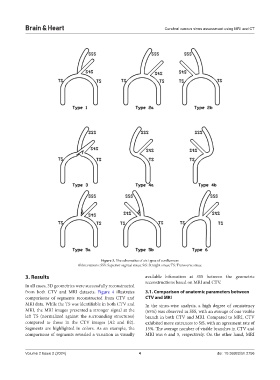
Figure 3. The schematics of six types of confluences
Abbreviations: SSS: Superior sagittal sinus; StS: Straight sinus; TS: Transverse sinus.
3. Results available bifurcation at SSS between the geometric
reconstructions based on MRI and CTV.
In all cases, 3D geometries were successfully reconstructed
from both CTV and MRI datasets. Figure 4 illustrates 3.1. Comparison of anatomic parameters between
comparisons of segments reconstructed from CTV and CTV and MRI
MRI data. While the TS was identifiable in both CTV and In the sinus-wise analysis, a high degree of consistency
MRI, the MRI images presented a stronger signal at the (65%) was observed in SSS, with an average of one visible
left TS (normalized against the surrounding structures) branch in both CTV and MRI. Compared to MRI, CTV
compared to those in the CTV images (A2 and B2). exhibited more entrances to StS, with an agreement rate of
Segments are highlighted in colors. As an example, the 15%. The average number of visible branches in CTV and
comparisons of segments revealed a variation in visually MRI was 6 and 5, respectively. On the other hand, MRI
Volume 2 Issue 2 (2024) 4 doi: 10.36922/bh.2756