
Page 50 - BH-2-2
P. 50
Brain & Heart Cerebral venous sinus assessment using MRI and CT
1. Introduction 2.2. Image acquisitions
Pulsatile tinnitus (PT) is often related to an underlying The MRI data were acquired using a 3.0T MRI unit
vascular abnormality, significantly affecting the patient’s (Ingenia, Philips Healthcare, Netherlands) equipped with
quality of life. Cerebral venous stenosis-induced PT a 16-channel head coil. The MRI examinations employed
1
has gained increasing recognition, with venous sinus a 3D phase-contrast technique using a gradient-echo
stenting (VSS) emerging as an effective treatment option sequence with the following parameters: field of view of
3
for symptom relief. Transverse sinus stenosis (TSS) is 173 × 173 × 192 mm , repetition time of 17 ms, echo time
2
a commonly observed vascular abnormality in patients of 6.2 ms, flip angle of 10°, velocity encoding of 15 cm/s,
with PT. The previous studies have highlighted the high bandwidth of 230 Hz/pixel, matrix size of 144 × 108 × 120,
3
diagnostic performance of both computed tomography and acquisition time of 2 min 15 s.
venography (CTV) and magnetic resonance venography CTV images were acquired using a 256-section
for anatomical analysis of venous sinuses. The choice CT scanner (Revolution, GE Healthcare, US) with the
4
of modality generally depends on availability, although following parameters: tube voltage of 100 kV, 25mAs (auto-
comparisons between modalities have been rarely assessed.
mAs), matrix of 512 × 512, collimation of 256×0.625 mm,
Anatomical assessment is widely conducted using rotation time of 0.5 s, pitch of 0.992:1, and administration
CTV and Phase-contrast magnetic resonance imaging of contrast media (iopamidol, Bracco Diagnostics, UK) at
(MRI). Computed tomography (CT) remains the primary a concentration of 370 mg iodine/ml, 1.5 ml/kg, injected
5,6
approach for differentiating the source of vascular-related at a rate of 5 ml/s. The average CT dose index (CTDI)
tinnitus, due to its high spatial resolution and consequent was 63.95 mGy, and the average total dose length product
high diagnostic accuracy. On the other hand, MRI (DLP) was 664.3 mGy∙cm.
3
has increasingly been applied for intracranial venous
assessment, offering advantages such as superior soft-tissue 2.3. Reconstruction of 3D geometry models
contrast without interference bone structures, as shown in The anatomies of the cases were reconstructed using
CT images. Therefore, MRI is considered a promising tool Mimics 19.0 (Materialise, Belgium). The region of interest
7-9
for analyzing the vasculature and the defects of surrounding was segmented based on the signal intensity distribution
tissues, including vascular interface, meningeal defects, and of MRI and CTV images. Consultation with clinical
related malformations. However, the previous studies technicians was conducted to improve the accuracy of
7,9
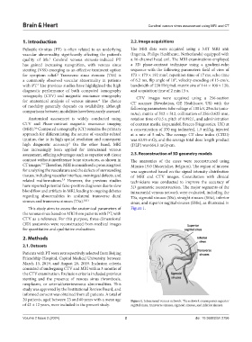
have reported potential false-positive diagnoses due to slow 3D geometric reconstruction. The major segments of the
blood flow and artifacts in MRI, leading to ongoing debates intracranial venous network were evaluated, including the
regarding abnormalities in unilateral transverse dural TSs, sigmoid sinuses (SSs), straight sinuses (StSs), inferior
sinuses and transverse sinuses (TSs). 10,11 sinus, and superior sagittal sinuses (SSSs), as illustrated in
This study aims to assess the anatomical parameters of Figure 1.
the venous sinus based on MRI from patients with PT, with
CTV as a reference. For this purpose, three-dimensional
(3D) anatomies were reconstructed from medical images
for quantitative and qualitative evaluations.
2. Methods
2.1. Datasets
Patients with PT were retrospectively selected from Beijing
Friendship Hospital, Capital Medical University, between
March 13, 2019, and August 26, 2019. Inclusion criteria
consisted of undergoing CTV and MRI within 3 months of
the CTV examination. Exclusion criteria included previous
stenting and the presence of venous sinus thrombosis,
neoplasms, or arterial/arteriovenous abnormalities. This
study was approved by the Institutional Review Board, and
informed consent was obtained from all patients. A total of
20 patients, aged between 23 and 60 years with a mean age Figure 1. Intracranial venous network. The network encompasses superior
of 42 ± 12 years, were included in the present study. sagittal sinus, transverse sinuses, sigmoid sinuses, and inferior sinuses.
Volume 2 Issue 2 (2024) 2 doi: 10.36922/bh.2756