
Page 94 - BH-2-4
P. 94
Brain & Heart Human DPSCs attenuated amyotrophic lateral sclerosis in mice
A B
C D E
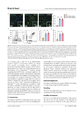
Figure 4. Microglia in SOD1-G93A mice showed no evident significant changes after treatment with hDPSCs. (A) Immunofluorescence images depicting
the amount of microglia in lumbosacral enlargement of the anterior horn of the spinal cord of WT and SOD1-G93A mice treated with vehicle or hDPSCs
on day 150. Scale bar = 50 µm. (B) Histogram illustrating the relative proportions of microglia in WT and SOD1-G93A mice treated with vehicle or
hDPSCs. n = 6 per group. (C) Flow cytometry analysis depicting the gating strategies used for microglia at day 150. (D) Histogram showing the percentage
of CD45 intermediate CD11b microglia (CD45 CD 11b+ ) in the brain of WT and SOD1-G93A mice treated with vehicle or hDPSCs on day 150. n = 4 in the WT
int
+
mice group, n = 6 in the vehicle-treated SOD1-G93A mice group, and n = 5 in the hDPSCs-treated SOD1-G93A mice group. (E) Histogram depicting the
percentage of CD45 intermediate CD11b microglia (CD45 CD11b ) in the spinal cord of WT and SOD1-G93A mice treated with vehicle or hDPSCs on day
+
int
+
150. n = 4 in the WT mice group, n = 6 in vehicle-treated SOD1-G93A mice group, and n = 5 in the hDPSCs-treated SOD1-G93A mice group. Data were
statistically analyzed using an unpaired t-test. Data were expressed as mean ± SEM. Notes: **p<0.01, ***p<0.001, and ****p<0.0001.
Abbreviations: hDPSCs: Human dental pulp stem cells; WT: Wild type; SEM: Standard error of mean.
the nanoscale plays a vital role in the differentiation cord atrophy and conserving neurons, demonstrating the
process of MSCs. 31,32 Furthermore, hDPSCs can secrete beneficial effects of hDPSCs infusion in ALS mice. This
some essential neurotrophic factors, including nerve evidence based on murine models needs further validation
growth factor proteins, BDNF, and glial cell line-derived by well-executed randomized controlled clinical trials.
neurotrophic factor, which can nourish damaged neurons Moreover, future optimized protocols for engineered
and promote neural regeneration. 33-35 Previous findings hDPSCs and the identification of reliable biomarkers to
suggesting that hDPSCs play a potential regulatory role predict therapeutic outcomes are essential steps toward the
in the inflammatory response during disease progression innovative use of hDPSCs for ALS treatment. Nevertheless,
were contradicted by the findings of the present study, as the use of hDPSCs as a therapeutic approach holds promise
the population of microglia or astrocytes in SOD1-G93A for future clinical interventions for patients with ALS.
mice exhibited no significant changes after the infusion of
hDPSCs. Studies have reported that extracellular vesicles Acknowledgments
36
released by MSCs induce functional modifications in
microglia. 37,38 Further investigation is required to clarify The authors thank Beijing Chengnuo Medical Technology
the impact of hDPSCs on glial cell features in ALS mice. Co., Ltd. for providing the hDPSCs injection.
Moreover, our study is limited by the fact that NeuN is Funding
not a specific marker for motor neurons. In addition, the
mechanism through which hDPSCs infusion increases the This work was supported in part by the National Science
levels of trophic factors that reduce the clinical symptoms Foundation of China (82122021).
of ALS mice remains unknown.
Conflict of interest
5. Conclusion Wei-Na Jin is an Editorial Board Member of this journal
The infusion of hDPSCs ameliorated clinical impairment but was not in any way involved in the editorial and
and prolonged the lifespan of ALS mice by retarding spinal peer-review process conducted for this paper, directly or
Volume 2 Issue 4 (2024) 7 doi: 10.36922/bh.3996