
Page 93 - BH-2-4
P. 93
Brain & Heart Human DPSCs attenuated amyotrophic lateral sclerosis in mice
A B
C D
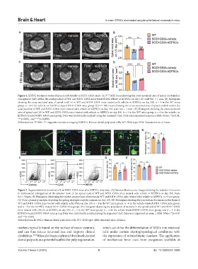
Figure 2. hDPSC treatment reduced spinal cord atrophy in SOD1-G93A mice. (A) 7-T MRI scans showing the cross-sectional area of spinal cord lumbar
enlargement (LE) within the axial position of WT and SOD1-G93A mice treated with vehicle or hDPSCs on day 120, scale bar = 1 mm. (B) Histogram
showing the cross-sectional area of spinal cord LE in WT and SOD1-G93A mice treated with vehicle or hDPSCs on day 120. n = 4 in the WT mice
group, n = 8 in the vehicle- or hDPSCs-treated SOD1-G93A mice group. (C) 7-T MRI scans showing the cross-sectional area of spinal cord LE within the
axial position of WT and SOD1-G93A mice treated with vehicle or hDPSCs on day 150, scale bar = 1 mm. (D) Histogram showing the cross-sectional
area of spinal cord LE in WT and SOD1-G93A mice treated with vehicle or hDPSCs on day 150. n = 4 in the WT mice group, n = 6 in the vehicle- or
hDPSCs-treated SOD1-G93A mice group. Data were statistically analyzed using the unpaired t-test. Data were expressed as mean ± SEM. Notes: **p<0.01,
***p<0.001, and ****p<0.0001.
Abbreviations: 7T MRI: 7T magnetic resonance imaging; hDPSCs: Human dental pulp stem cells; WT: Wild type; SEM: Standard error of mean.
A B
C D E
Figure 3. Augmentation of neuronal cells in SOD1-G93A mice after hDPSCs treatment. (A) Immunofluorescence images showing the number of neurons
in lumbosacral enlargement of the anterior horn of the spinal cord of WT and SOD1-G93A mice treated with vehicle or hDPSCs on day 150. Scale
bar = 50 µm. (B) Histogram illustrating the relative proportions of neurons in WT and SOD1-G93A mice treated with vehicle or hDPSCs. n = 6 per group.
(C) Flow cytometry analysis depicting the gating strategies used for neurons on day 150. (D) Histogram showing the population of neurons in the brain of
WT and SOD1-G93A mice treated with vehicle or hDPSCs on day 150. n = 4 in the WT mice group, n = 6 in the vehicle-treated SOD1-G93A mice group,
and n = 5 in the hDPSCs-treated SOD1-G93A mice group. (E) Histogram depicting the population of neurons in the spinal cord of WT and SOD1-G93A
mice treated with vehicle or hDPSCs on day 150. n = 4 in the WT mice group, n = 6 in the vehicle-treated SOD1-G93A mice group, and n = 5 in the
hDPSCs-treated SOD1-G93A mice group. Data were statistically analyzed using the unpaired t-test. Data were expressed as mean ± SEM. Notes: **p<0.01
and ***p<0.001.
Abbreviations: hDPSCs: Human dental pulp stem cells; WT: Wild type; SEM: Standard error of mean.
markers typically found on the surface of motor neurons, which can drive the differentiation of MSCs into neuronal
and can thus rescue neuronal loss and improve clinical cells under certain electrophysiological conditions with
disabilities. 29,30 It has also been confirmed that decellularized the expression of odontoblastic markers. The application
dental pulp acts as a potential scaffold for pulp regeneration, of mechanical force cues from exogenous scaffolds at
Volume 2 Issue 4 (2024) 6 doi: 10.36922/bh.3996