
Page 64 - BH-3-1
P. 64
Brain & Heart Thrombectomy for stroke after cardiac surgery
revealed a mitral valve prolapse with massive mitral NIHSS score at 24 h had improved to 6. The patient was
regurgitation. The ejection fraction was normal. The discharged, and her mRS score at 3 months was 1.
patient underwent mechanical mitral valve replacement
and tricuspid valvuloplasty. Five days after valve surgery, 3.3. Case 3
she experienced right hemiparesis, with an initial NIHSS A 50-year-old man with valvular disease underwent
score of 16. CTA revealed a left internal carotid artery mitral valve replacement and tricuspid valvuloplasty. The
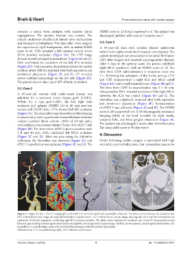
(ICA) terminal occlusion (Figure 2A). The CTP image patient developed new-onset atrial onset atrial fibrillation
showed several salvageable penumbrae (Figure 2B and C). (AF) after surgery and received anticoagulation therapy.
DSA confirmed the occlusion of the left ICA terminal After 4 days in the general ward, the patient exhibited
(Figure 2D). Unfortunately, the perforator from the middle right MCA syndrome, with an NIHSS score of 14. The
cerebral artery (MCA) ruptured after both aspiration and time from LKW administration to symptom onset was
stentriever placement (Figure 2E and F). CT revealed 1 h. Following the activation of the stroke service, CTA
severe cerebral hemorrhage on the left side (Figure 2G). and CTP demonstrated a right ICA and MCA cutoff
The patient died on day 1 post-MT of brain herniation. (Figure 4A) and a small mismatch area (Figure 4B and C).
3.2. Case 2 The time from LKW to recanalization was 5 h 20 min.
Intraoperative DSA revealed occlusion of the right MCA;
A 65‐year‐old woman with triple-vessel disease was however, the ICA was patent (Figure 4D and E). The
admitted for a coronary artery bypass graft (CABG). thrombus was completely removed after both aspiration
Within the 4 days post‐CABG, she had right limb and stentriever placement (Figure 4F). Recanalization
weakness and aphasia (NIHSS 16) at 40 min post‐last of eTICI 3 was achieved (Figure 4G and H). The NIHSS
known well (LKW) time. CTA showed left M1 occlusion score at 24 h improved to 8. A 10-day magnetic resonance
(Figure 3A). The penumbra map showed favorable imaging
characteristics, with a predicted irreversible brain ischemia imaging (MRI) of the head revealed the right insula,
volume (cerebral blood volume <30%) of 1.9 mL and a temporal lobe, and basal ganglia infarctions (Figure 4I).
total ischemic brain tissue volume (Tmax >6 s) of 217.7 mL The patient was discharged 2 weeks after thrombectomy.
(Figure 3B). The times from LKW to groin puncture were The mean mRS score at 90 days was 0.
3 h and 40 min. DSA confirmed left MCA occlusion 4. Discussion
(Figure 3C and D). After one pass using the Solumbra
technique, the thrombus was removed (Figure 3E) and Stroke following cardiac surgery is associated with high
eTICI 3 reperfusion was achieved (Figure 3F and G). The mortality and morbidity rates. Our consecutive case series
A B D E G
C
F
Figure 2. Images of case 1. (A) CT angiography with a left ICA terminal cutoff and reasonable collaterals. The white arrow indicates the occlusion site.
(B) Cerebral blood flow image showing left hemisphere hypoperfusion. (C) Cerebral blood volume image showing that both cerebral hemispheres are
equivalent. (D) Initial angiogram confirming right ICA terminal occlusion. The white arrow indicates the occlusion site. (E and F) Anteroposterior and
lateral images showing contrast agent extravasation during MT, indicating cerebral hemorrhage. The blue arrows indicate contrast agent extravasation. (G)
Immediate CT scan showing a large area of cerebral hemorrhage in the left cerebral hemisphere.
Abbreviations: CT: Computed tomography; ICA: Internal carotid artery.
Volume 3 Issue 1 (2025) 4 doi: 10.36922/bh.4141