
Page 65 - BH-3-1
P. 65
Brain & Heart Thrombectomy for stroke after cardiac surgery
A B C D
F G
E
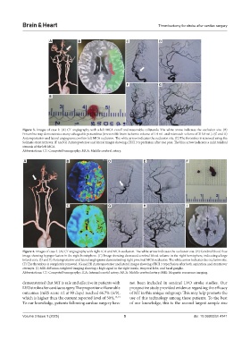
Figure 3. Images of case 2. (A) CT angiography with a left MCA cutoff and reasonable collaterals. The white arrow indicates the occlusion site. (B)
Penumbra map demonstrates many salvageable penumbras (irreversible brain ischemic volume of 1.9 mL and mismatch volume of 215.8 mL). (C and D)
Anteroposterior and lateral angiograms confirm left MCA occlusion. The white arrow indicates the occlusion site. (E) The thrombus is removed using the
Solitaire stent retriever. (F and G) Anteroposterior and lateral images showing eTICI 3 reperfusion after one pass. The blue arrow indicates a mild residual
stenosis of the left MCA.
Abbreviations: CT: Computed tomography; MCA: Middle cerebral artery.
A B D E F
C G H I
Figure 4. Images of case 3. (A) CT angiography with right ICA and MCA occlusion. The white arrow indicates the occlusion site. (B) Cerebral blood flow
image showing hypoperfusion in the right hemisphere. (C) Image showing decreased cerebral blood volume in the right hemisphere, indicating a large
infarct core. (D and E) Anteroposterior and lateral angiograms demonstrating right proximal MCA occlusion. The white arrow indicates the occlusion site.
(F) The thrombus is completely removed. (G and H) Anteroposterior and lateral images showing eTICI 3 reperfusion after both aspiration and stentriever
attempts. (I) MRI diffusion-weighted imaging showing a high signal in the right insula, temporal lobe, and basal ganglia.
Abbreviations: CT: Computed tomography; ICA: Internal carotid artery; MCA: Middle cerebral artery; MRI: Magnetic resonance imaging.
demonstrated that MT is safe and effective in patients with not been included in sentinel LVO stroke studies. Our
LVO stroke after cardiac surgery. The proportion of favorable prospective study provided evidence regarding the efficacy
outcomes (mRS score ≤2 at 90 days) reached 66.7% (6/9), of MT in this unique subgroup. This may help promote the
which is higher than the current reported level of 50%. 13,14 use of this technology among these patients. To the best
To our knowledge, patients following cardiac surgery have of our knowledge, this is the second largest sample size
Volume 3 Issue 1 (2025) 5 doi: 10.36922/bh.4141