
Page 9 - BH-3-1
P. 9
Brain & Heart TAVR in low gradient aortic stenosis
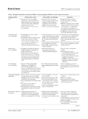
Table 1. Strengths, limitations, and clinical utility of various imaging modalities in aortic stenosis assessment
Imaging modality Objectives/Key concept Clinical utility and advantages Limitations
TTE • Assessment of valve morphology; • The most effective initial non- • Suboptimal acoustic windows
quantification of the severity of AS by invasive imaging modality for the • Limited visualization of the distal
measuring the peak jet velocity, mean evaluation of AS, enabling the thoracic aorta
pressure gradient, AVA, and Doppler visualization of the aortic valve • Difficulty in assessing valve parameters
velocities across the valve leaflets and any abnormalities in patients with low-flow, low-gradient
• LVOT assessment, LV function, and Crucial for accurate AVA calculations AS, the presence of LV dysfunction,
hemodynamics concomitant aortic regurgitation, or
discordant hemodynamic measurements
• Calcification causes artifacts that
obscure the underlying structures
Dobutamine stress • To distinguish true-severe AS from • Guides management: True-severe AS • Possible adverse effects of dobutamine,
echocardiography pseudo-severe AS requires valve replacement, whereas such as ventricular arrhythmias and
• No alteration in the AVA with increased pseudo-severe AS may benefit from hypertension, in select patients
cardiac output indicates a severely the treatment of underlying heart • Time-consuming and requires clinical
stenotic valve. An increase in AVA of failure expertise
>1 cm with an increased flow rate is • Useful in patients with low-gradient • The presence or absence of flow reserve
2
observed in pseudo-severe AS AS and impaired LV function does not affect management, as both
Determine the absence of flow reserve • Predicts high operative mortality benefit from AVR
and poor prognosis
Exercise stress • Although not routinely indicated, this • Better risk stratification in • Cannot be used in symptomatic
echocardiography modality is helpful in asymptomatic asymptomatic patients than patients
patients with severe AS conventional echocardiography • Contraindications include:
• An increase in the mean transaortic 1. An established indication for AVR
pressure gradient by ≥18 – 20 mmHg 2. Uncontrolled hypertension
during exercise is linked to an elevated 3. Symptomatic or hemodynamically
risk of cardiac death, development of significant arrhythmias
spontaneous symptoms, and need for 4. Inability to conduct the test due to
AVR orthopedic limitations or global
disabilities
Transesophageal • Adjunct to TTE, providing direct • Pre-TAVR assessment of patients 2D echocardiography may result in
echocardiography visualization of the leaflets and • High-quality imaging comparable compromising the TAVI implant owing
subvalvular abnormalities in the to CT to the elliptical nature of the LVOT and
presence of suboptimal acoustic windows annulus
To determine accurate measurements of
the aortic valve annulus, leaflet mobility,
and calcification
Computed tomography • Superior spatial and temporal resolution • Assess the suitability of the native • Not effective in determining the extent
angiography in visualizing the aortic valve leaflets, valve for TAVR of fibrosis
including assessment of their mobility, • Select the most appropriate THV • Exposure to radiation
calcification, and morphology • Determine the optimal vascular • Risks associated with contrast
• To accurately measure the aortic annulus access route for TAVR to assist in administration, especially in patients
size procedural planning and mitigate with renal impairment
• To evaluate the vessel diameter, vascular complications
tortuosity, and calcification • Guide revascularization strategies
• To evaluate the coronary artery anatomy before or during TAVR
and identify the presence of obstructive
CAD
Multidetector computed • Semiquantitative assessment of • Highly accurate and reproducible • Does not provide hemodynamic
tomography calcification of the aortic valve by the information that is independent of information such as transvalvular
Agatston score and calcium densities the flow and hemodynamics pressure gradients or the presence of
(calcium score adjusted for LVOT area) • Does not require the administration regurgitation
of contrast • Frequently produces motion artifacts in
patients with higher resting heart rates.
• Radiation exposure.
• Can underestimate the severity in
select individuals.
(Cont’d...)
Volume 3 Issue 1 (2025) 3 doi: 10.36922/bh.4017