
Page 95 - BH-3-1
P. 95
Brain & Heart A neurological association of bicuspid aortic valve
A B C
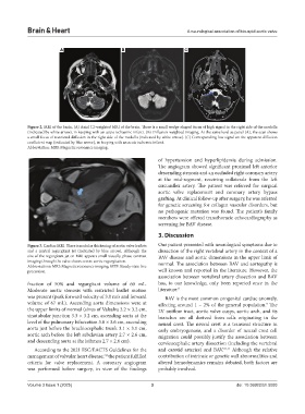
Figure 2. MRI of the brain. (A) Axial T2-weighted MRI of the brain. There is a small wedge-shaped focus of high signal in the right side of the medulla
(indicated by white arrow), in keeping with an acute ischaemic infarct. (B) Diffusion-weighted imaging. At the same level as panel (A), the scan shows
a small focus of restricted diffusion in the right side of the medulla (indicated by white arrow). (C) Corresponding low signal on the apparent diffusion
coefficient map (indicated by blue arrow), in keeping with an acute ischemic infarct.
Abbreviation: MRI: Magnetic resonance imaging.
of hypertension and hyperlipidemia during admission.
The angiogram showed significant proximal left anterior
descending stenosis and an occluded right coronary artery
at the mid-segment, receiving collaterals from the left
circumflex artery. The patient was referred for surgical
aortic valve replacement and coronary artery bypass
grafting. At clinical follow-up after surgery, he was referred
for genetic screening for collagen vascular disorders, but
no pathogenic mutation was found. The patient’s family
members were offered transthoracic echocardiography as
screening for BAV disease.
3. Discussion
Figure 3. Cardiac MRI. There is nodular thickening of aortic valve leaflets Our patient presented with neurological symptoms due to
and a central regurgitant jet (indicated by blue arrow). Although the dissection of the right vertebral artery in the context of a
size of the regurgitant jet on MRI appears small visually, phase-contrast BAV disease and aortic dimensions in the upper limit of
imaging through the valve shows severe aortic regurgitation. normal. The association between BAV and aortopathy is
Abbreviations: MRI: Magnetic resonance imaging; SSFP: Steady-state free
precession. well known and reported in the literature. However, the
association between vertebral artery dissection and BAV
fraction of 50% and regurgitant volume of 60 mL. has, to our knowledge, only been reported once in the
Moderate aortic stenosis with restricted leaflet motion literature. 6
was present (peak forward velocity of 3.0 m/s and forward BAV is the most common congenital cardiac anomaly,
volume of 67 mL). Ascending aorta dimensions were at affecting around 1 – 2% of the general population. The
9
the upper limits of normal (sinus of Valsalva 3.2 × 3.2 cm, LV outflow tract, aortic valve cusps, aortic arch, and its
sinotubular junction 3.3 × 3.2 cm, ascending aorta at the branches are all derived from cells originating in the
level of the pulmonary bifurcation 3.6 × 3.6 cm, ascending neural crest. The neural crest is a transient structure in
aorta just before the brachiocephalic trunk 3.1 × 3.1 cm, early embryogenesis, and a disorder of neural crest cell
aortic arch before the left subclavian artery 2.7 × 2.6 cm, migration could possibly justify the association between
and descending aorta at the isthmus 2.7 × 2.6 cm). cervicocephalic artery dissection (including the vertebral
According to the 2021 ESC/EACTS Guidelines for the and carotid arteries) and BAV. 10,11 Although the relative
management of valvular heart disease, the patient fulfilled contribution of intrinsic or genetic wall abnormalities and
7,8
criteria for valve replacement. A coronary angiogram altered hemodynamics remains debated, both factors are
was performed before surgery, in view of the findings probably involved.
Volume 3 Issue 1 (2025) 3 doi: 10.36922/bh.5093