
Page 94 - BH-3-1
P. 94
Brain & Heart A neurological association of bicuspid aortic valve
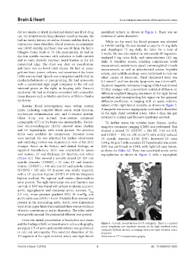
did not smoke or drink alcohol and denied any illicit drug medullary infarct, as shown in Figure 1. There was no
use. He denied recent long-distance travel or trauma. He evidence of aortic dissection.
had no family history of cardiac disease, sudden death, or While on the ward, his blood pressure was elevated
connective tissue disorders. Blood pressure on admission to 150/90 mmHg. He was started on aspirin 75 mg daily
was 140/85 mmHg and heart rate was 60 bpm. He had a and clopidogrel 75 mg daily, the latter for a total of
Glasgow Coma Scale of 15. On examination, he had an 3 weeks. He was also started on atorvastatin 80 mg nocte,
ejection systolic murmur with radiation to both carotids enalapril 5 mg twice daily, and spironolactone 12.5 mg
and an early diastolic murmur, heard loudest at the left daily. A vasculitis screen, entailing complement levels
parasternal edge. The chest was clear on auscultation, measurement, autoimmune panel, immunoglobulin levels
and there was no lower limb edema. He had an ataxic measurement, as well as antiretroviral screen, hepatitis
gait and tone, power, reflexes, and sensation of the lower screen, and syphilis serology were performed to rule out
limbs were normal. Speech was unimpaired and he had no other causes of dissection. Total cholesterol level was
dysdiadochokinesia or past-pointing. He had anisocoria 6.2 mmol/L and low-density lipoprotein was 4.4 mmol/L.
with a constricted right pupil compared to the left and Inpatient magnetic resonance imaging of the head showed
minimal ptosis on the right, in keeping with Horner’s T2-flair changes with concomitant restricted diffusion on
syndrome. He had no features consistent with connective diffusion-weighted imaging sequences of the right lateral
tissue diseases such as Marfan syndrome or Ehlers–Danlos medullary and corresponding low signal on the apparent
syndrome. diffusion coefficient, in keeping with an acute ischemic
Routine blood investigations were within normal infarct of the right lateral medulla, as shown in Figure 2.
limits, including complete blood count, renal function, A magnetic resonance angiography confirmed a dissection
erythrocyte sedimentation rate, and C-reactive protein. of the right distal vertebral artery. After 3 days, his gait
Chest X-ray was normal. Non-contrast computed returned to normal and Horner’s syndrome resolved.
tomography (CT) of the brain was unremarkable. Twelve- To further assess the valvular heart disease, cardiac
lead electrocardiogram (ECG) showed sinus rhythm magnetic resonance imaging (CMR) was performed. This
and LV hypertrophy with strain pattern. No previous showed a dilated LV (LVEDV = 280 ML [139 mL/m ],
2
ECGs were available for comparison. Troponin levels and LVESV = 162 mL (80 mL/m ]) with mildly reduced
2
were normal. He was admitted for further observation LV systolic function (LVEF 42%). Increased LV mass
and cardiology review was requested in view of the ECG (194 g, 96 g/m ) with eccentric LV hypertrophy was noted.
2
changes. Based on the history and clinical findings, an BAV was confirmed on CMR, with right-left cusp fusion,
inpatient transthoracic ECG was conducted to assess as shown in Video A2. There was associated severe aortic
wall motion, LV wall thickness, LV function, and valves regurgitation, as shown in Figure 3, with a regurgitant
(Video A1). This showed a severely dilated LV (LV end
systolic diameter [LVESD] = 51 mm; LV end diastolic
volume [LVEDV] = 198 mL; and LV end systolic volume
[LVESV] = 102 mL). LV function was mildly impaired,
with a LV ejection fraction (LVEF) of 45% by Simpson’s
biplane method. No regional wall motion abnormalities
were present. The right ventricular size and function was
normal. A BAV was found with at least moderate eccentric
aortic regurgitation and moderate aortic stenosis, V
max
3.3 m/s, mean pressure gradient (PG) 30 mmHg, and
aortic valve area (AVA) 1.4 cm . Diastolic flow reversal was
2
present in the descending aorta. Aortic root dimensions
were at the upper limit of normal and there was no evidence
of aortic coarctation or aortic dissection. The other valves
were grossly normal. No pericardial effusion was present.
Given the initial presentation of headaches and ataxia
and the finding of BAV on transthoracic echocardiography, Figure 1. Coronal reconstruction of CT aortogram. There is a marked
an urgent CT of aorta and carotid arteries was performed mural irregularity and moderate stenosis of the right vertebral artery
(indicated by black arrows), in keeping with acute right vertebral artery
to rule out arteriopathy. This unveiled dissection of the dissection.
V3 segment of the right vertebral artery and right lateral Abbreviation: CT: Computed tomography.
Volume 3 Issue 1 (2025) 2 doi: 10.36922/bh.5093