
Page 49 - GPD-2-2
P. 49
Gene & Protein in Disease Dunaliella salina & myocardial ischemia-reperfusion injury
A
B C D
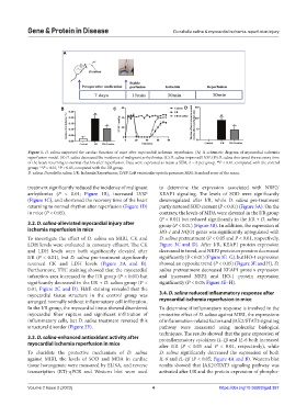
Figure 1. D. salina improved the cardiac function of mice after myocardial ischemia reperfusion. (A) A schematic diagram of myocardial ischemia
reperfusion model. (B) D. salina decreased the incidence of malignant arrhythmias. (C) D. salina improved LVSP. (D) D. salina shortened the recovery time
of the heart returning to normal rhythm after reperfusion. Data were expressed as mean ± SEM, n = 8 per group. P < 0.01, compared with the control
##
group; **P < 0.01, *P < 0.05, compared with the I/R group.
D. salina: Dunaliella salina; I/R: Ischemia/Reperfusion; LVSP: Left ventricular systolic pressure; SEM: Standard error of the mean.
treatment significantly reduced the incidence of malignant to determine the expression associated with NRF2/
arrhythmias (P < 0.01; Figure 1B), increased LVSP KEAP1 signaling. The levels of SOD were significantly
(Figure 1C), and shortened the recovery time of the heart downregulated after I/R, while D. salina pre-treatment
returning to normal rhythm after reperfusion (Figure 1D) partly restored SOD content (P < 0.01) (Figure 3A). On the
in mice (P < 0.05). contrary, the levels of MDA were elevated in the I/R group
(P < 0.01) but reduced significantly in the I/R + D. salina
3.2. D. salina-alleviated myocardial injury after group (P < 0.01) (Figure 3B). In addition, the expression of
ischemia reperfusion in mice HO-1 and NQO1 genes was significantly upregulated with
To investigate the effect of D. salina on MIRI, CK and D. salina pretreatment (P < 0.05 and P < 0.01, respectively,
LDH levels were evaluated in coronary effluent. The CK Figure 3C and D). After I/R, KEAP1 protein expression
and LDH levels were both significantly elevated after decreased in trend, and NRF2 protein expression decreased
I/R (P < 0.01), but D. salina pre-treatment significantly significantly (P < 0.01) (Figure 3E–G), but HO-1 expression
reversed CK and LDH levels (Figure 2A and B). showed an opposite trend (P < 0.05) (Figure 3E and H). D.
Furthermore, TTC staining showed that the myocardial salina pretreatment decreased KEAP1 protein expression
infarction area increased in the I/R group (P < 0.01) but and increased NRF2 and HO-1 protein expression
significantly decreased in the I/R + D. salina group (P < significantly (P < 0.05; Figure 3E–H).
0.01, Figure 2C and D). H&E staining revealed that the
myocardial tissue structure in the control group was 3.4. D. salina reduced inflammatory response after
arranged normally without inflammatory cell infiltration. myocardial ischemia reperfusion in mice
In the I/R group, the myocardial tissue showed disordered To determine if inflammatory response is involved in the
myocardial fiber rupture and significant infiltration of protective effect of D. salina against MIRI, the expressions
inflammatory cells, but D. salina treatment reversed this of inflammation-related factors and JAK2/STAT3 signaling
structural disorder (Figure 2E). pathway were measured using molecular biological
techniques. The results showed that the gene expression of
3.3. D. salina-enhanced antioxidant activity after proinflammatory cytokines IL-1β and IL-6 both increased
myocardial ischemia reperfusion in mice after I/R (P < 0.05 and P < 0.01, respectively), while
To elucidate the protective mechanism of D. salina D. salina significantly decreased the expression of both
against MIRI, the levels of SOD and MDA in cardiac IL-6 and IL-1β (P < 0.05; Figure 4A and B). Western blot
tissue homogenate were measured by ELISA, and reverse results showed that JAK2/STAT3 signaling pathway was
transcription (RT)-qPCR and Western blot were used activated after I/R and the protein expression of phospho-
Volume 2 Issue 2 (2023) 4 https://doi.org/10.36922/gpd.387