
Page 51 - GPD-2-2
P. 51
Gene & Protein in Disease Dunaliella salina & myocardial ischemia-reperfusion injury
A B
C D E
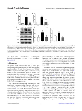
Figure 4. D. salina reduced inflammatory response after myocardial ischemia reperfusion in mice. Gene expression of inflammatory cytokines (A) IL-1β
and (B) IL-6. (C) Representative Western blot images of JAK2/STAT3 signaling pathway. Quantity analysis of (D) p-JAK2 and (E) p-STAT3. Data were
##
expressed as mean ± SEM, n = 8 per group. P < 0.01, P < 0.05, compared with the control group; **P < 0.01, *P < 0.05, compared with the I/R group.
#
D. salina: Dunaliella salina; GAPDH: Glyceraldehyde-3-phosphate dehydrogenase; I/R: Ischemia/Reperfusion; IL: Interleukin; JAK2: Janus kinase 2;
mRNA: messenger RNA; p-JAK2: phospho-JAK2; p-STAT3: phospho-STAT3; SEM: Standard error of the mean; STAT3: Signal transducer and activator
of transcription 3.
JAK2 (p-JAK2) and p-STAT3 in the I/R + D. salina group preconditioning of D. salina could reduce myocardial
decreased significantly (P < 0.05 and P < 0.01, respectively, infarction area. In addition, H&E staining showed that
Figure 4C–E). D. salina preconditioning reduced myocardial damage
at the morphological level. Taken together, these results
4. Discussion suggest that D. salina pretreatment could protect the heart
The present study demonstrated that D. salina pre- from MIRI.
treatment provides protection against MIRI and the effect Enormous ROS can consume endogenous antioxidant
may be mediated through the enhancement of KEAP1/ substances and enzymes, cause peroxidation of the lipid
[21]
NRF2 endogenous antioxidant system and the weakening bilayer of the cell membrane, and lead to cell injury .
[22]
of JAK2/STAT3 inflammatory signaling pathway. The SOD can effectively scavenge ROS , while MDA, a
results indicated the potential of D. salina as a promising marker of lipid peroxidation, indicates the degree of
candidate drug in the prevention of MIRI. The present lipid peroxidation [23,24] . In the D. salina pre-treatment
study is the first study to show that the protective effect group, SOD activity increased and the content of MDA
of D. salina on MIRI may be related to KEAP1/NRF2 and significantly reduced in the myocardial tissue, indicating
JAK2/STAT3 signaling pathways. that D. salina can significantly reduce the oxidative stress
level. The KEAP1/NRF2 signaling pathway is an important
The Langendorff perfused heart is a classical in vitro endogenous antioxidant defense system . NRF2 regulates
[25]
model used to examine cardiac function, such as LVSP the transcription of antioxidant-related genes by binding to
and heart rate, without the influence of complex internal ARE . NRF2 and KEAP1 bind together in the cytoplasm
[26]
environment . The results showed that D. salina pre- under normal physiological conditions. When stimulated,
[19]
treatment could increase LVSP and decrease the incidence KEAP1 degrades, and NRF2 is released from the
of malignant arrhythmias within 30 min of reperfusion. cytoplasm. Then, NRF2 nuclear translocation occurs, and
In addition, CK and LDH are often used as indicators of NRF2 binds with MAF to form heterodimers . D. salina
[27]
myocardial injury . The results showed that the levels of administration upregulated NRF2 expression and its
[20]
CK and LDH decreased significantly after pretreatment downstream antioxidative enzymes, such as HO-1 and
with D. salina. TTC staining results also showed that the NQO1, which may be related to the degradation of KEAP1.
Volume 2 Issue 2 (2023) 6 https://doi.org/10.36922/gpd.387