
Page 88 - GPD-2-2
P. 88
Gene & Protein in Disease Verteporfin therapy for triple-negative breast cancer
A B C D
E
F G
H I
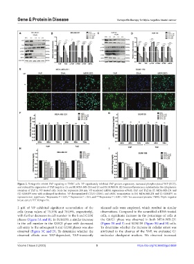
Figure 2. Verteporfin inhibit YAP signaling in TNBC cells. VP significantly inhibited YAP protein expression, increased phosphorylated YAP (S127),
and reduced the expression of YAP targets in (A and B) MDA-MB-231 and (C and D) SUM 159. (E) Immunofluorescence substantiates the cytoplasmic
retention of YAP in VP-treated cells. Scale bar represents 200 µm. VP unaltered mRNA expressions of both YAP and TAZ in (F) MDA-MB-231 and
(G) SUM159 even with prolonged incubation. VP downregulated CTGF, CYR61, and cMYC transcription in (H) MDA-MB-231 and (I) SUM159. ns
represents non-significant, *Represents P < 0.05, **Represents P < 0.01, and ***Represents P < 0.001. YAP: Yes-associated protein; TNBC: Triple-negative
breast cancer; VP: Verteporfin.
2 µM of VP exhibited significant accumulation of the silenced cells were employed, which resulted in similar
cells (mean values of 75.35% and 78.19%, respectively), observations. Compared to the scrambled siRNA-treated
with further decreases in cell number in the S and G2/M cells, a significant increase in the percentage of cells at
phases (Figure 5A and B). In SUM159, a similar increase the G0/G1 phase was observed in both MDA-MB-231
in the cell number in the G0/G1 phase with decreased (Figure 5E and F) and SUM159 (Figure 5G and H) cells.
cell entry to the subsequent S and G2/M phases was also To determine whether the increase in cellular arrest was
observed (Figure 5C and D). To determine whether the attributed to the absence of the YAP, we evaluated G1
observed effects were YAP-dependent, YAP-transiently molecular checkpoint markers. We observed increased
Volume 2 Issue 2 (2023) 5 https://doi.org/10.36922/gpd.0658