
Page 34 - GPD-2-4
P. 34
Gene & Protein in Disease Cyanine and cancer therapy
of cancer at different stages involves alterations in
mitochondrial function [42-45] , making mitochondria one
of the key targeted organelles for anti-cancer therapy
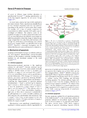
(Figure 1) .
[46]
In recent years, cyanine has been widely employed in
anti-tumor research. These compounds are classified based
on their chemical composition into Cy3, Cy5, and Cy7,
depending on the number of carbon atoms they contain.
For example, Cy7, a type of cyanine compound, has
demonstrated the ability to produce ROS under a specific
wavelength of radiation. This property opens up the
possibility of using Cy7 to induce apoptosis in tumor cells.
Cy7, as a novel fluorescent dye with anti-cancer properties,
holds the potential to reduce the dosage of chemotherapy
drugs, mitigate their toxic side effects, and treat tumor cells
through the combination of chemotherapy, near-infrared Figure 1. The role of mitochondria in apoptosis. Mechanistically,
mitochondria serve as the primary sites for producing ROS during
fluorescence imaging (NIRF), and photothermal therapy apoptosis. These ROS can induce changes in intracellular MOMP by
[47]
(PTT) . Therefore, a thorough investigation into the acting on the mitochondrial membrane, which is regulated by proteins in
relationship between cyanine and apoptosis is imperative. the Bcl-2 family. Subsequently, the release of Cyto C from mitochondria
activates cysteine cascades, leading to cell apoptosis.
2. Mechanism of apoptosis Abbreviations: AKT: Protein kinase B; AMPK: Adenosine
5’-monophosphate(AMP)-activated protein kinase; BAD: Bcl-xl/Bcl-2
Apoptosis can be initiated through two different pathways: associated death promoter; BAX: Bcl-2 associated X protein; Bcl-2: B cell
the Bcl-2-regulated mitochondrial cystatin-dependent lymphoma 2; BIM: Bcl-2 interacting mediator of cell death; DISC: Death-
pathway and the exogenous apoptotic signaling pathway inducing signaling complex; DR5: Death receptor 5; FAS: Apoptosis
stimulating fragment; GSH: glutathione (L-gamma-Glutamyl-L-cysteinyl-
mediated by cell membrane proteins of the death glycine); JNK1: Recombinant c-Jun N-terminal kinase 1; MOMP:
receptor [22,48,49] . Mitochondrial outer membrane permeabilization; mTOR: Mammalian
target of rapamycin; NAC: N-Acetyl-L-cysteine; NADPH: Nicotinamide
2.1. Intrinsic apoptosis adenine dinucleotide phosphate; P53: Protein 53; PKM2: Active Pyruvate
kinase isozymes M2; PTEN: Phosphatase and tensin homolog deleted on
Mitochondrial-mediated apoptosis is the significant chromosome ten; ROS: Reactive oxygen species.
pathway for cell death . Intrinsic pathways can be
[50]
induced by various intracellular stress stimuli, eventually interaction of multiple proteins from the members of the
leading to MOMP, release of Cyto C, formation and pro-apoptotic Bcl-2 family, such as Bcl-2, BAX, and the
activation of caspase-9, and increased ROS levels [8,26] . Bcl-2 antagonist/killer 1 (BAK) . MOMP is essential for
[57]
Cells sense intracellular stressors such as growth factors, inducing the release of apoptosis-inducing factor (AIF) ,
[39]
nutrient deficiencies, DNA damage, UV radiation, and which subsequently translocates freely into the cytoplasm
hypoxia, and induce apoptosis by initiating MOMP . or nucleus, causing DNA breakage and ultimately
[6]
During MOMP, Cyto C is released from mitochondria , mediating apoptosis. The intrinsic pathway begins with
[51]
promoting the oligomerization of apoptotic protease the release of Cyto C into the cytoplasm, followed by its
activating factor 1 (Apaf-1), and forming the heptamer. activation by pro-apoptotic proteins . The release of free
[58]
Apaf-1 contains a, which combines with pro-caspase-9 Cyto C into the cytoplasm is a crucial requirement for
to form the apoptosome, a large complex of Cyto C, triggering apoptosis and is regarded as a point of no return
Apaf-1, and pro-caspase-9 [52-56] . This pathway relies on in cell death, occurring independently of cystathionin
the release of Cyto C from mitochondria, requiring a activation (Figure 2) [54,59,60] .
high ratio of BAX (Bcl-2 associated X protein)/Bcl-2
(B cell lymphoma 2) to create favorable conditions for 2.2. Extrinsic apoptosis
BAX insertion into mitochondria and subsequent Cyto Extrinsic apoptosis, mediated by cell membrane proteins
C release . Cyto C then recruits caspase-9 to form the of the death receptor , is the second major pathway of
[10]
[27]
apoptosome complex. Ultimately, activation of caspase-3 apoptosis, initiated by extracellular signals originating
initiates the caspase cascade . from the immune system or the external environment .
[26]
[8]
Mitochondrial outer membrane permeabilization is The tumor necrosis factor (TNF) family receptors,
significant in intrinsic apoptosis, which is regulated by the consisting of apoptosis stimulating fragment (FAS)-R,
Volume 2 Issue 4 (2023) 3 https://doi.org/10.36922/gpd.2486