
Page 97 - GTM-3-2
P. 97
Global Translational Medicine Targeted detection in Barrett’s neoplasia
is time-consuming and limited by sampling error and is EGFR and HER2 was topically administered to the Barrett’s
therefore not widely practiced by community physicians. segment. Wide-field NIR fluorescence images were
6
Pre-malignant lesions (LGD and HGD) are often flat in captured using the multi-modal scanning fiber endoscope
appearance and patchy in distribution. Other methods, such (mmSFE) as an imaging accessory. 12,13 Fluorescence images
7
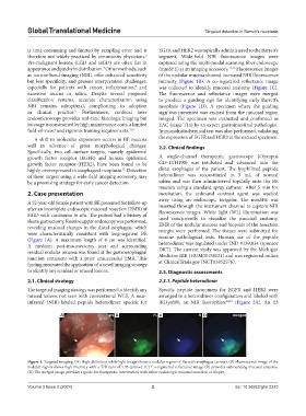
as narrow-band imaging (NBI), offer enhanced sensitivity of the nodular mucosa showed increased NIR fluorescence
but less specificity, and present interpretation challenges, intensity (Figure 1B). A co-registered reflectance image
especially for patients with cancer, inflammation, and was collected to identify mucosal anatomy (Figure 1C).
8
excessive mucus or saliva. Despite several proposed The fluorescence and reflectance images were merged
classification systems, accurate characterization using to produce a guiding sign for identifying early Barrett’s
NBI remains suboptimal, complicating its adoption neoplasia (Figure 1D). A specimen where the guiding
in clinical practice. Furthermore, confocal laser sign was presented was excised from the mucosal region
9
endomicroscopy provides real-time histologic imaging but imaged. The specimen was evaluated and confirmed as
its usage is constrained by high maintenance costs, a limited EAC (stage T1a) by an expert gastrointestinal pathologist.
field-of-view, and rigorous training requirements. 7,10 Immunohistochemical test was also performed, validating
8
A shift in molecular expression occurs in BE mucosa the expression of EGFR and HER2 in the excised specimen.
well in advance of gross morphological changes. 2.2. Clinical findings
Specifically, two cell-surface targets, namely epidermal
growth factor receptor (EGFR) and human epidermal A single-channel therapeutic gastroscope (Olympus
growth factor receptor (HER2), have been found to be GIF-1TH190) was intubated and advanced into the
highly overexpressed in esophageal neoplasia. Detection distal esophagus of the patient. The lyophilized peptide
11
of these targets using a wide-field imaging accessory may heterodimer was reconstituted in 5 mL of normal
be a promising strategy for early cancer detection. saline and was then administered topically onto the BE
mucosa using a standard spray catheter. After 5 min for
2. Case presentation incubation, the unbound contrast agent was washed
away using an endoscopic irrigator. The mmSFE was
A 52-year-old female patient with BE presented for follow-up inserted through the instrument channel to capture NIR
after an incomplete endoscopic mucosal resection (EMR) of fluorescence images. White light (WL) illumination was
HGD with carcinoma in situ. The patient had a history of used concurrently to visualize the mucosal anatomy.
sleeve gastrectomy. Routine upper endoscopy was performed, EMR of the nodular mucosa and biopsies of the resection
revealing mucosal changes in the distal esophagus, which margin were performed. The tissues were submitted for
were characteristically consistent with long-segment BE routine pathological tests. Human use of the peptide
(Figure 1A). A maximum length of 4 cm was identified. heterodimer was regulated under IND #139,834 (sponsor
A medium post-mucosectomy scar and surrounding DKT). The current study was approved by the Michigan
residual nodular mucosa was found at the gastroesophageal Medicine IRB (HUM00158121) and was registered online
junction consistent with a prior unsuccessful EMR. This
finding motivated the application of a novel imaging strategy at ClinicalTrials.gov (NCT03852576).
to identify any residual or missed lesions. 2.3. Diagnostic assessments
2.1. Clinical strategy 2.3.1. Peptide heterodimer
The targeted imaging strategy was performed to identify any Specific peptide monomers for EGFR and HER2 were
missed lesions not seen with conventional WLE. A near- arranged in a heterodimer configuration and labeled with
infrared (NIR)-labeled peptide heterodimer specific for IRDye800, an NIR fluorophore 14,15 (Figure 2A). An E3
A B C D
Figure 1. Targeted imaging. (A) High-definition white light image shows a nodular region of Barrett’s esophagus (arrow). (B) Fluorescence image of the
nodular region shows high intensity with a T/B ratio of 1.93 (arrow). (C) Co-registered reflectance image (R) provides surrounding mucosal anatomy.
(D) The merged image provides a guide for therapeutic intervention with either endoscopic mucosal resection or biopsy.
Volume 3 Issue 2 (2024) 2 doi: 10.36922/gtm.2223