
Page 14 - GTM-3-3
P. 14
Global Translational Medicine Influence of estrogen on RV mitochondria in PH
GRP30) (Figure 1). 49,50 ERα and ERβ share a great deal of 4. Influence of estrogen on mitochondrial
similarities although they have significant differences. ERα function
is composed of an amino-terminal transcription control
domain (AF-1), which is its main region of interaction E2 affects the mitochondria both indirectly through
with regulatory binding proteins; however, ERβ does not targeting the nucleus or directly by regulating the expression
have a strong AF-1 domain within its amino terminus. of mitochondrial genes. Both ERα and ERβ have been
51
Instead, ERβ contains a repressor domain that modulates detected within the mitochondria, with ERβ accounting
61,62
ERα activity. Meanwhile, GPER is unrelated to the ERs as the main receptor (Figure 2). Mitochondrial ERs are
51
but does indeed, mimic ER signaling. ERα and ERβ encoded by the same genes that encode nuclear ERα and
52
reside in the nucleus, mitochondria, and cytoplasm, 53,54 ERβ as knock-out of ERα and ERβ demonstrates a complete
63
while GPER is expressed in the plasma membrane as absence of mitochondrial ER in mice. The localization of
well as the endoplasmic reticulum. Sex differences these receptors within the mitochondria varies depending
53
have been detected in ERα protein expression, with ERα on the cell type. The classical estrogen signaling mechanism
expression significantly higher in female cardiomyocytes is when E2 passes through the plasma membrane and
than in males. Meanwhile, ERβ protein expression in binds directly with intracellular ERα and ERβ in the
cardiomyocytes is similar in both males and females. 55,56 cytoplasm (Figure 2). This binding triggers receptor
phosphorylation and dimerization, in which this newly
E2 will diffuse into the cell where it will locate the formed complex translocates into the nucleus where it
nuclear ER and trigger receptor dimerization. These dimers binds to the chromatin at ERE sequences, enhancer regions,
then interact directly with specific DNA sequences, known and 3’-untranslated regions of target genes (Figure 2). 57,64
as estrogen response elements (ERE), which transactivate ERα and ERβ can also be phosphorylated and activated
gene expression. Alternatively, E2 can interact indirectly in a ligand-independent manner. 49,57 There are over 70,000
through the tethering of other DNA transcription factors, EREs within the mouse and human genomes. These
65
leading to the recruitment of activator proteins. 57,58 GPER nuclear effects can then influence mitochondrial DNA
will be activated through the classical G protein-coupled (mtDNA) gene transcription and function. For instance,
receptor mechanism. The genomic effects mediated by the E2 can regulate nuclear respiratory factor 1 (NRF1), which
ERs (ERα, ERβ) occur over hours to days while the non- promotes the transcription of mitochondrial transcription
66
genomic effects mediated by GPER occur rapidly within factor A (TFAM) that targets the mtDNA genes. E2 can
seconds to minutes. 59,60 also promote the upregulation of peroxisome proliferator-
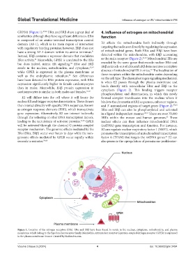
Figure 1. Location of the estrogen receptors (ERs). ERα and ERβ have been found to reside in the nucleus, cytoplasm, mitochondria, and plasma
membrane, which belong to the type I nuclear receptor family. Meanwhile, extranuclear receptor G protein-coupled estrogen receptor (GPER) is expressed
in the plasma membrane. Source: Created by BioRender.com.
Volume 3 Issue 3 (2024) 4 doi: 10.36922/gtm.2494