
Page 44 - GTM-3-3
P. 44
Global Translational Medicine Ocular changes in Alzheimer’s disease
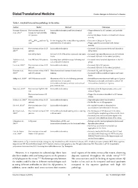
Table 1. Amyloid beta and tau pathology in the retina
Author Model Method Outcomes
Koronyo-Hamaoui Post-mortem retina of Immunohistochemistry and histochemical • Plaques detected in AD patients, and probable
et al., 2011 31 human AD and probable staining cases
AD patients • Retinal Aβ plaque burden correlated with disease
severity.
APP SWE /PS1 and non-Tg In vivo imaging of the retina following systemic • Detection of plaque in Tg mice
ΔE9
mice administration of curcumin • Plaque quantity accumulated with disease
severity.
Koronyo et al., Post-mortem retina of AD Immunohistochemistry • Increased Aβ immunoreactivity and deposits in
2017 22 patients AD patients
Live AD patients SLO and OCT of the retina upon oral curcumin • Increased curcumin fluorescence in AD patients
intake • Increased Aβ levels in the retinal periphery.
Tadokoro et al., Live MCI and AD patients Scanning laser ophthalmoscopy following oral • Increased retinal amyloid deposition in the AD
2021 32 curcumin administration group
Lee et al., 2020 33 Post-mortem retina of AD Immunohistochemistry • Increased Aβ in AD patients
patients • Increased Aβ levels in the retinal periphery.
Koronyo et al., Post-mortem retina of MCI Histochemical and immunohistochemical • Increased Aβ and Aβ oligomers
42
2023 35 and AD patients staining • Increased levels in peripheral regions and inner
layers.
Sidiqi et al., 2020 23 APP/PS1 mouse model Fluorescence SLO in vivo following systemic • Retinal fluorescence increased with age in Tg mice
administration of curcumin • Fluorescence strongly correlated to cortical Aβ
Immunohistochemistry on brain and retina of immunohistochemistry in Tg mice.
post-mortem mice
Tsai et al., 2014 36 Post-mortem TgF344-AD Immunohistochemistry • Aβ detected in the hippocampus, cortex, and
rats retina of Tg rats.
Post-mortem human AD • Plaque-like structures identified in AD human
retina retinas.
Schön et al., 2012 37 Post-mortem retina of AD Immunohistochemistry • Hyperphosphorylated tau identified
patients • No fibrillar tau or Aβ aggregates.
Ho et al., 2014 38 Post-mortem eyes of AD Immunohistochemistry • No amyloid deposits or abnormal tau
patients accumulations were detected in the eyes.
den Haan et al., Post-mortem retinas of AD Immunohistochemical staining and co-staining • No Aβ plaques or NT detected in the retina
2018 39 patients with curcumin resembling the morphology of those in the brain
• Increased p-tau immunoreactive signal in the
inner and outer plexiform layers of the retina.
Chiasseu et al., 3×Tg mice Western blots, qPCR, and • Epitope-specific tau hyperphosphorylation and
2017 40 immunohistochemistry on the brain and retina hypophosphorylation
• Retinal tau accumulation preceded tau
aggregation in the brain.
Hart de Ruyter Post-mortem retina and Immunohistochemistry • Retinal p-tau Ser202/Thr205 and Thr217 load
et al., 2023 41 brains of AD patients correlated with Braak stage for NFTs and p-tau
SER202/Thr205 levels in the hippocampus and
cortical brain regions.
Abbreviations: 3×Tg: 3-month-old triple transgenic; Aβ: Amyloid beta; AD: Alzheimer’s disease; APP: Amyloid precursor protein; MCI: Mild cognitive
impairment; NT: Neurofibrillary tangle; OCT: Optical coherence tomography; p-tau: Phosphorylated tau; PS1: Presenilin 1; qPCR: Quantitative
polymerase chain reaction; SLO: Scanning laser ophthalmoscope; Tg: Transgenic.
However, it is important to acknowledge there have small regions of the retina versus a flat mount, observing
also been studies with conflicting reports on the presence deposits scattered throughout the whole retina. 31,36,37,39
of Aβ plaques in the retina. 37-39 The heterogeneity between The cross-sections could be looking at regions where Aβ
the studies could be due to different methodologies such burden is low, such as the temporal and nasal quadrants
as using different antibodies to label the Aβ proteins. In compared to the superior quadrant with high AD
addition, some studies used cross-sections that examine pathology.
Volume 3 Issue 3 (2024) 5 doi: 10.36922/gtm.4094